In the past, bacteriophage λ DNA purification was largely achieved through procedures that involved Cesium chloride gradients. However, these methods had a low recovery rate and a high chance of the presence of contaminants. Modern bacteriophage λ DNA purification methods give high yield and provide DNA free from contamination (Pickard, 2009).
We can also purify bacteriophage λ DNA from liquid lysates. Bacteriophage λ shows better growth in plate lysates as compared to small liquid cultures. Therefore, the given method is suitable only for those strains which are robust such as; λZAP, λZipLox, λgt11, and λgt10.
Alternative Protocols
Various methods have been described for purifying bacteriophage λ from plate lysates and liquid cultures. However, almost all of these methods are minor variations or expansions of the basic protocols. The most commonly encountered problems are:
- The inability of DNA to get cleaved by restriction endonucleases
One can resolve the first problem by varying the actual and relative amounts of bacteriophage to bacterial cell ratio. The yield of bacteriophage DNA largely depends on the bacteriophage to bacterial cell ratio. Therefore, while working with a different strain of E. coli, it is better to prepare a set of reactions with different phage to cell ratios. Therefore, their absolute and relative amounts are compared, and the resulting liquid lysate is used to prepare λ DNA.
The second problem arises due to polysaccharide inhibitors present in bacteria or agar. If the contaminants are from agar, it is better to use agarose instead. However, if the problem remains, the bacteriophage particles need to get purified by any of the following procedures.
- One variation of this method is to first remove polyanions from the lysate by using DEAE cellulose. Proteinase K and EDTA are then added to extract DNA from phage particles. Later on, hexadecyltrimethylammonium bromide (CTAB) is added to precipitate bacteriophage DNA.
- Another variation is to use a protonated form of the resin during chromatography. The negatively charged virus particles bind to Mg+2 ions present in low concentrations in the resin and are then eluted in buffers with high Mg+2 concentrations. Proteinase K and SDS are then used to digest the virus and release DNA. This DNA is precipitated with isopropanol.
- One can use commercially available kits to purify DNA from phage particles as well. The contaminants are removed by nuclease, DNA is precipitated in PEG/resin by centrifugation, and then DNA is purified by another resin.
By following any of the two methods (liquid cultures and plate lysate method), we can purify approximately 5ꭒg DNA from 5×10¹⁰ infectious particles. With the innovations in molecular biology, researchers have come up with procedures that can yield up to 23ml of 1011 PFUs. Although these procedures are lengthy and may take from 16 to 21 days, they can play a significant role in personalized phage therapy (Luong et al., 2020).
Materials
Buffers, Media and Solutions
- Chloroform
- Ethanol
- SM
- High Salt Buffer
- 20mM Tris-Chloride (pH 7.4)
- 1.0M NaCl
- 1mM EDTA (pH 8.0)
- Low Salt Buffer
- 20mM Tris-Chloride (pH 7.4)
- 0.2M NaCl
- 1mM EDTA (pH 8.0)
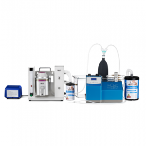
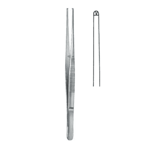
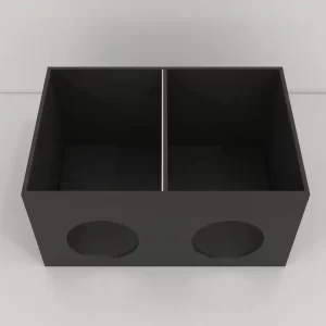
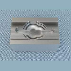
Other Equipment
Bacterial and Viral Strains
Inoculate an E. coli strain in 25ml of NZCYM medium and store overnight at 30oC. Measure O.D. Calculate the number of cells per ml using formula 1O.D.600 = 1×10⁹ cells/ml. The 30oC incubation temperature ensures that the cell growth does not reach saturation to minimize cell debris. If phages are added to a saturated culture, they attach to MalB protein in cell debris and cause “non-productive infection.”
- Recombinant bacteriophage λ, grown as single well-isolated plaque on a lawn of bacteria
Methods
- In a sterile microfuge tube, add 1ml of SM and 50ml of chloroform. Pick a single well-isolated plaque with a Pasteur pipette and suspend it in this tube.
- Incubate the microfuge tube at 4oC for 4 to 6 hours and let the bacteriophage particles diffuse from the top agarose.
- Mix 0.1ml of overnight bacterial culture with 0.5ml of phage suspension in a 25ml tube. Store this infected culture at 37oC for 15 minutes.
- Now add 4ml of NZCYM medium to the infected culture and store it at 37oC for about 9 hours with vigorous shaking. At this step, the culture must become clear, and a little bit of cell debris should be visible.
- Add 1ml of chloroform to the tube and incubate for additional 15 minutes under the same conditions. Shift the lysate to a 5ml centrifuge tube and centrifuge at 2600rpm for 10 minutes at 4oC.
- Pour the supernatant into a fresh tube. Now centrifuge at 5800rpm for 10 minutes at 4oC to remove the cell debris. Separate a small aliquot of lysate at this step and store it at 4oC over chloroform as bacteriophage stock.
- Dispense 10ml 2:1 slurry of a DEAE: cellulose resin in a clean microfuge tube. Centrifuge the tube at 2000rpm (500g) for 5 minutes at room temperature. The centrifugation will cause the resin to sediment. Remove the supernatant and incubate the tube containing the pellet on ice.
- Resuspend the resin pellet into bacteriophage λ supernatant. Place the centrifuge tube on a rocking platform for 3 minutes to facilitate the absorption of phage particles to the resin.
- Centrifuge the resultant slurry at 5400rpm (4000g) for 5 minutes. Pour the supernatant into a fresh tube and centrifuge once again under the same conditions. Discard the pellet.
- Again, transfer the supernatant to a fresh centrifuge tube and subject the supernatant (containing bacteriophage particles) to phenol: chloroform extraction once.
- Now, shift the bacteriophage λ DNA containing an aqueous phase to a new tube and add an equal volume of isopropanol. Incubate the mixture at -70oC for 10 minutes.
- Centrifuge this mixture at 12000rpm for 20 minutes at 4oC. Collect the precipitated bacteriophage DNA.
- Discard isopropanol from the tube and let the DNA pellet get air-dried.
- Dissolve the DNA in 2ml of a low salt buffer.
- Purify the bacteriophage DNA by chromatography as described in the previous protocol.
- Mix eluate with 1ml of ethanol and place the mixture on ice for 20 minutes. Centrifuge to precipitate out DNA, discard the supernatant.
- Wash the pellet with 0.5ml of 70% ethanol, discard the supernatant and let ethanol evaporate.
- Resuspend the damp pellet in a drop of TE (pH 8.0).
To analyze the DNA, perform agarose gel electrophoresis. If necessary, perform restriction digestion before electrophoresis.
Precautions
- Sometimes, problems might arise due to the contamination of bacterial DNA or RNA. In this case, treat the culture with DNase I before chromatography. For this, add DNase I to a final concentration of 10ng/ml before step 5. The sample’s viscosity is decreased by this treatment which might enhance the bacterial DNA or RNA removal.
- In case the lysis is incomplete or does not occur in step 4, pre-warm an equal volume of NZCYM medium and add it to the culture. Incubate for an additional 2 to 3 hours at 37oC with vigorous shaking. Consequently, lysis will be complete, and this will produce a higher yield of DNA.
- Do not vortex the pellet in the final step. Redissolve the pellet by gently tapping on the sides of the tube. If the DNA still does not dissolve, incubate the tube again for 15 minutes at 50oC.
Summary
- Apart from plate lysates, liquid cultures can also be used to purify bacteriophages. However, this method is suitable for robust strains only.
- The poor yield of DNA and DNA’s inability to get cleaved by restriction endonucleases are the most common problems encountered in this procedure.
- The yield of DNA is improved by taking into account the bacteriophage to bacterial cell ratio.
- Several variations in the protocol are adopted to overcome the problem of the inability to get cleaved. These methods include CTAB treatment, treatment with protonated resin before chromatography, and the use of commercially available kits.
References
- Luong, T., Salabarria, A. C., Edwards, R. A., & Roach, D. R. (2020). Standardized bacteriophage purification for personalized phage therapy. Nature Protocols, 15(9), 2867-2890.
- Pickard, D. J. J. (2009). Preparation of bacteriophage lysates and pure DNA. In Bacteriophages (pp. 3-9). Humana Press.
- Sambrook, J., & Russell, D. W. (2006). Rapid Analysis of Bacteriophage λ Isolates: Purification of λ DNA from Liquid Cultures. Cold Spring Harbor Protocols, 2006(1), pdb-prot3985.