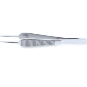
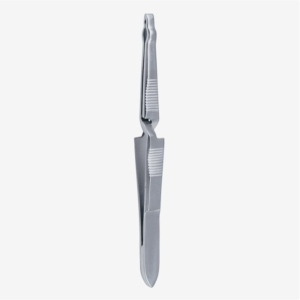
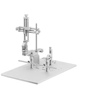
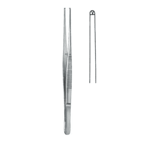
Are you looking for Slice Electrophysiology devices for your lab?

Electrophysiology Lab: Definition

Electrophysiology labs are centers that provide care for people having arrhythmias (heart rhythm disorders) or neural disorders.[1] Thus, the lab is split into cardiac electrophysiology and neural electrophysiology labs.

Electrophysiology labs perform electrophysiology studies. In a heart condition, the heart’s electrical activity is recorded using a catheter inserted through a vein or artery.[3] This is done using a special X-ray “movie” called fluoroscopy. It helps in understanding the origin of disease, its cause, and suitable treatment for the diseased patients.[4]

In case of neural challenges (especially those associated with the brain, retina, and spine), procedures like the patch clamp technique and sharp microelectrodes are used to study the activity of astrocytes, and neurons, and communication between different neurons.[14] 

Electrophysiology studies are also done if you have any symptoms like weakness, palpitation, fainting, or dizziness to check if it’s caused by any heart condition or interruptions in nerve communications.[4]

The electrophysiology lab is equipped with high-throughput equipment and tools to perform procedures like ablation, cardiac mapping, fast-scan cyclic voltammetry, patch-clamp, radiofrequency ablation, automated ECG diagnostics, optogenetics, and pacemaker installation. Additionally, it’s done to determine if the given medicine for heart disorders or neural problems is effective for patients.

In this article, we will explore more on cardiac electrophysiology labs, the procedures performed in these labs, the devices used, and their applications in different medicinal or research areas.

What Procedures Are Done in a Cardiac Electrophysiology Lab?

The electrophysiology procedure is a test performed to assess and determine the electrical activity of the heart and diagnose any abnormal conditions, such as arrhythmia.[5]

In arrhythmia, the heart’s signals that coordinate its beats do not function properly; they either beat too slow (a condition known as bradycardia) or too fast (tachycardia).[2]

A range of procedures are used in such labs to study, diagnose, or treat these conditions. Some of them are given below:

Cardiac ablation

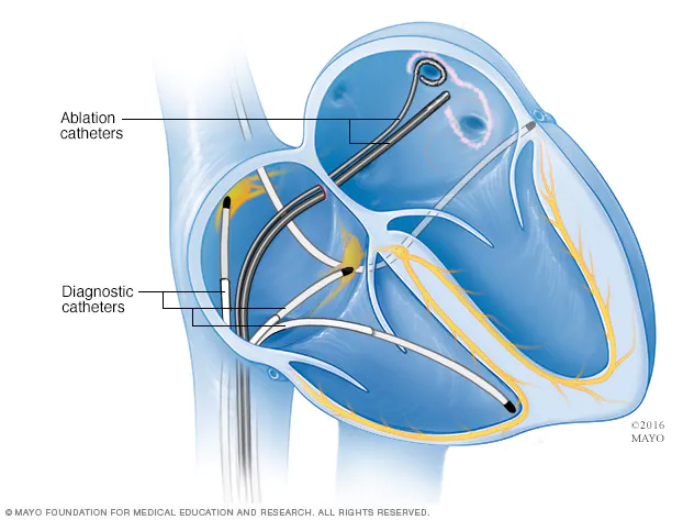
The technique utilizes heat or cold to create tiny scars in the heart. It’s done by inserting a catheter (thin, flexible tubes) through the arteries and veins.[6] The sensors present on the catheter’s tip destroy the tissue and create ablation. Thus, irregular electrical signals are blocked and a normal heartbeat is restored. The procedure is mainly performed during cardiac surgery.

Illustration of cardiac ablation procedure with catheter placement inside the heart

Figure: An illustration of cardiac ablation procedure with catheter placement inside the heart.[6]

Often it’s one of the first treatments recommended to patients with heart rhythm problems, while other times, it’s used to treat diseases for which other treatments or medicines aren’t working.

Implantable devices

These are biological devices implanted in a patient’s body to serve a specific function during surgery and clinical procedures.

In electrophysiology labs, an implantable device is a pacemaker placed in a patient’s chest using surgical procedures to control their heartbeat.[7]  Its types are single chamber pacemakers, dual chamber pacemakers, and biventricular pacemakers.[7]

A pacemaker is recommended when a person has a slow heartbeat after a surgery or heart attack to recover from the abnormal heart condition.

Implantable cardioverter-defibrillators (ICDs)

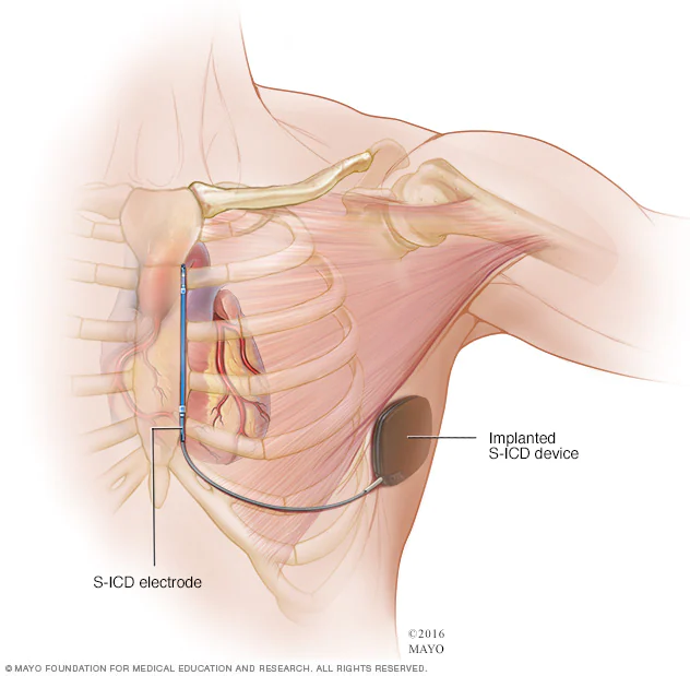
It’s a small device powered by a battery installed in a patient’s chest suffering from arrhythmia to detect and stop irregular heartbeats.[8] The device constantly monitors heartbeats and delivers electric shocks to restore regular heartbeats as needed.

It’s required when one has ventricular tachycardia or ventricular fibrillation condition or has a weak heart muscle. Its types include a traditional ICD and subcutaneous ICD (S-ICD).

It’s used either when a person has survived cardiac arrest, coronary artery disease, or certain irregular heart rhythm.

An illustration of implantation of a Subcutaneous implantable cardioverter-defibrillator (S-ICD) device just underneath the armpit on the side of the chest

Figure: An illustration of implantation of a Subcutaneous implantable cardioverter-defibrillator (S-ICD) device just underneath the armpit on the side of the chest.[8]

Radiofrequency ablation

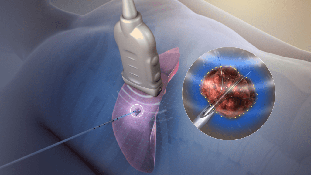
It’s also known as radiofrequency neurotomy. The technique uses the current (created by radiofrequency waves) to generate heat in the targeted diseased tissue. It destroys the nerve tissues in the area and hinders the pain signals sent to the brain.[9]

The technique is extensively used to end chronic pain like heart rhythm disorders, tumors, peripheral nerve pain, or joint pain. It’s widely used in cardiac arrhythmia to recurrent atrial fibrillation (AF), atrial tachycardia, atrial flutter (Afl), supraventricular tachycardia (SVT), and some other types of ventricular arrhythmia.[9]

An illustration of electric current (produced by radiofrequency) ablating tumor tissues

Figure: An illustration of electric current (produced by radiofrequency) ablating tumor tissues.[9]

Cryoablation

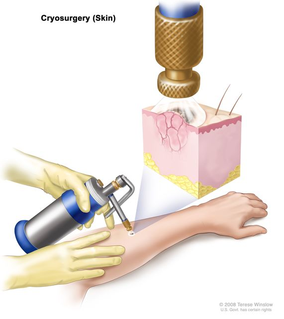
The procedure involves using an extremely cold instrument or substance to destroy abnormal or damaged tissues. Liquid nitrogen, liquid nitrous oxide, compressed argon gas, and carbon dioxide are commonly used substances in the procedure.[10]

The procedure has application in cryosurgery and cryotherapy to treat conditions like arrhythmia, cancer, skin abnormalities, and cervix precancerous cells.[10]

An illustration of cryoablation procedure on skin

Figure: An illustration of cryoablation procedure on skin.[10]

Cardiac mapping

Here, an anesthetic is given to the patient and an incision is made in the patient’s thigh or neck. A catheter is then introduced into the vein or artery through the incision.[11]

The catheter contains electrodes at its end, which gather data on electrical signals flowering through the patient’s heart. The technique is widely used to treat heart rhythm disorders such as arrhythmia.[11]

What Devices Are Used in a Cardiac Electrophysiology Lab?

Cardiac electrophysiology labs are equipped with high-throughput equipment and devices to perform the surgical procedures in the labs. Some of these pieces of equipment include:[12]

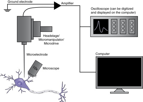
  1. Microelectrode: It is an electrode consisting of small diameter metal wires or fibers (5-100 µm) either sealed in metal or glass bodies. It detects the changes in the extracellular field caused by the current flows across the morphology of the closest neuron and other nearby cells. They are essential for electrophysiological recordings.[12]
  2. Headstage: It’s the central hub that facilitates the connection between electronic equipment and tissue preparation.
  3. Microdrive: Using a microdrive, the microelectrode can be lowered or raised to a particular depth in tissue whenever required.
  4. Amplifier: It’s used to amplify the weak signals received from microelectrodes. The signal is then transferred to a computer or oscilloscope for data visualization and interpretation.[12]
  5. Oscilloscope: It displays the data obtained from the amplifier.
  6. Loudspeaker system: Any changes in the voltage over time can be heard by using this instrument. Moreover, it’s an effective method while locating neurons, because neurons in different states produce different sounds. For Example, neurons in an active state produce a popping sound.[12]
  7. Microscope: The equipment is necessary for all extracellular and intracellular recording, especially while applying physiology or patch clamp techniques.
  8. Faraday Cage: It’s equipment made from conductive tools. It’s used to block or eliminate noses from sensitive electrical recordings.

Other than these instruments, some others used in the electrophysiology labs include X-ray tables, camera and television screens, temperature control devices, heart monitors, vibration isolation systems, and computers.[12]

Some critical components of the electrophysiology labs

Figure: Some critical components of the electrophysiology labs.[12]

What Are the Applications of Cardiac Electrophysiology Labs?

Electrophysiology labs have extensive application in disease diagnosis or treatments, it’s widely used in cardiology labs to study heart rhythm disorders, find their root cause, and potential solutions for the treatment of the disease.

Some of the uses include:

  • Cardiac electrophysiology labs are used to perform ablation procedures for patients suffering from atrial fibrillation.[13]
  • The lab is used by patients requiring a pacemaker for normal heart rhythm or to control their heartbeat.
  • Patients suffering from arrhythmia get a defibrillator inserted into the heart to protect them from life-threatening arrhythmic conditions. [13]
  • Cardiac electrophysiology labs are equipped with the industry’s most advanced biplane procedure rooms with advanced recording and analysis equipment to perform inpatient electrophysiology studies.[1]
  • For diagnosing and treating a spectrum of diseases, including Atrial flutter, Sudden cardiac arrest, heart failure requiring implanted device-based therapy, atrial fibrillation, and Syncope (fainting), Bradycardia, Supraventricular tachycardia, and Ventricular tachycardia.[1]
  • Cardiac electrophysiology labs also have applications in research studies. It’s used to conduct research on heart rhythm disorders and locate neurons in active or inactive states.[1]

Conclusion

Cardiac electrophysiology labs are facilities equipped with high-throughput equipment to provide care for patients with heart rhythm disorders. The lab utilizes electrical procedures to detect the signals obtained from the heart and determine the heartbeat.

Most commonly, a catheter having electrodes at the end is inserted inside the patient’s heart through arteries and veins, which note the beats obtained from the heart and send them to an oscilloscope for data visualization.

The most common instruments used in the lab include microelectrode, X-ray table, microscope, headstage, amplifier, and vibration isolation systems.

If you need a temperature control device to help maintain specimen temperature for electrophysiological experiments, check out our visual patching & imaging chamber.

References

  1. Electrophysiology Laboratory. Retrieved from https://www.mayoclinic.org/departments-centers/electrophysiology-laboratory/overview/ovc-20442531
  2. Heart arrhythmia. Retrieved from https://www.mayoclinic.org/diseases-conditions/heart-arrhythmia/symptoms-causes/syc-20350668#:~:text=A%20heart%20arrhythmia%20(uh%2DRITH,slow%20(bradycardia)%20or%20irregularly
  3. Electrophysiology study. Retrieved from https://en.wikipedia.org/wiki/Electrophysiology_study
  4. Electrophysiological Studies. Retrieved from https://www.hopkinsmedicine.org/health/treatment-tests-and-therapies/electrophysiological-studies
  5. Electrophysiology Procedure. Retrieved from https://www.ucsfhealth.org/education/electrophysiology-procedure
  6. Cardiac ablation. Retrieved from https://www.mayoclinic.org/tests-procedures/cardiac-ablation/about/pac-20384993
  7. Implantable cardioverter-defibrillators (ICDs). Retrieved from https://www.mayoclinic.org/tests-procedures/implantable-cardioverter-defibrillators/about/pac-20384692
  8. Radiofrequency ablation. Retrieved from https://en.wikipedia.org/wiki/Radiofrequency_ablation#/media/File:Radiofrequency_ablation.jpg
  9. Cryoablation. Retrieved from https://www.cancer.gov/publications/dictionaries/cancer-terms/def/cryoablation
  10. Cryoablation. Retrieved from https://my.clevelandclinic.org/health/treatments/16903-cryoablation
  11. Cardiac Mapping. Retrieved from https://www.leehealth.org/our-services/cardiology/cardiac-mapping
  12. Carter, M., & Shieh, J. (2015). Electrophysiology. Guide to Research Techniques in Neuroscience, 89–115. doi:10.1016/b978-0-12-800511-8.00004-6
  13. What is an electrophysiology lab? Retrieved from https://www.matherhospital.org/care-treatment/cardiac-cath-lab-electrophysiology-lab/
  14. Introduction To Electrophysiology. Retrieved from. https://www.photometrics.com/learn/electrophysiology/introduction-to-electrophysiology