$439.00
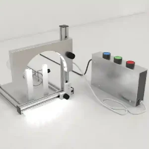
Integrated Horizontal Electrophoresis System (Electrophoresis Tank + blue light transilluminator)
Horizontal electrophoresis tanks are generally used for the analysis and size separation of nucleic acids, i.e., DNA and RNA.
The horizontal electrophoresis system is made of acrylic and comprises two buffer chambers, each containing an electrode. The apparatus consists of a casting tray, horizontal electrophoresis tank (also known as a submarine tank), gel combs, electrodes, and a power supply. Conduct Science offers two different models of horizontal electrophoresis tanks. Each model has different sizes of casting trays and gel combs.
ConductScience offers Electrophoresis Tanks.
Power pack sold separately, please see here.

Servicebio is a company that specializes in providing high-quality products and services for biomedical research and diagnostics.


SVL-2 integrated horizontal electrophoresis instrument is mainly used for rapid agarose electrophoresis separation experiments of small amounts of DNA and RNA samples.
Specifications | SB-SVL2 |
|---|---|
Specifications and Dimensions (LXWXH) | 310 X 150 X120mm |
Gel Tray Specifications (LXW) | Large gel 120mm X 120mm; Width gel 60mm X120mm; Long gel 120mm X 60mm; Small gel 60mm X 60mm |
Specimen Comb Specifications | 1.0mm 25/11 teeth 1.5mm 13/6 teeth 2.0mm 3/2 tooth |
Blue light wavelength | 470nm |
Buffer capacity | 260mL |
Weight | 1kg |
Item | Product | Quantity |
|---|---|---|
1 | Horizontal Electrophoresis apparatus | 1 set |
2 | Gel maker | 1 |
3 | 1.0mm 25-tooth/11-tooth sample comb | 4 |
4 | 1.5mm 13-tooth/6-tooth sample comb | 1 |
5 | 2.0mm 3-tooth/2-tooth sample comb | 1 |
6 | 60mm X 60mm Gel Tray | 2 |
7 | 60mm X 120mm Gel Tray | 1 |
8 | 120mmX60mm gel tray | 1 |
9 | 120mmX 120mm gel tray | 1 |
10 | Blue light transilluminator | 1 set |
1. The gel maker mold is integrally formed, which can make four different sizes ofgel blocks.
2. Standard blue light transilluminator, with SPW-6S electrophoresis power supply, can directly observe the band during the electrophoresis process.
3. The transparent upper cover has a hole design, which is convenient for heat dissipation and observation.
4. Card slot limit function, accurate operation.
5. The electrode holder and electrode head are detachable, which is convenient for cleaning and maintenance.
Horizontal electrophoresis tanks are generally used for the analysis and size separation of nucleic acids, i.e., DNA and RNA. However, it can also be used for the analysis of protein complexes. In a horizontal electrophoresis system, the gel matrix is cast horizontally and submerged in a running buffer within the electrophoresis tank. The tank is divided into two compartments separated by agarose gel.
Horizontal electrophoresis tanks contain an anode at one end and a cathode at another. A charge gradient is created as the electric current flows through the ionic running buffer. The purpose of the running buffer is to cool the gel, which gets heated up as the charge is applied. The occasional recirculation of running buffer prevents the formation of a pH gradient. Unlike vertical electrophoresis tanks, the two chambers in horizontal systems are connected by running buffers and cannot be used with a discontinuous buffer system.
Horizontal gel electrophoresis most commonly employs agarose gel. Agarose gels have larger pores, about 100-500nm in diameter. These gels help determine the size of the nucleic acids accurately based on their mobility through the gel. Acrylamide gel cannot be usually used for horizontal systems as the gel gets exposed to atmospheric oxygen in these systems, and oxygen prevents acrylamide polymerization, thereby interfering with gel formation. However, some modern electrophoresis protocols use polyacrylamide gel in horizontal systems.
The horizontal electrophoresis system is made of acrylic and comprises two buffer chambers, each containing an electrode. The apparatus consists of a casting tray, horizontal electrophoresis tank (also known as a submarine tank), gel combs, electrodes, and a power supply. Conduct Science offers two different models of horizontal electrophoresis tanks. Each model has different sizes of casting trays and gel combs. Power pack not included, please see here.
The underlying protocol is followed for a typical agarose gel electrophoresis for the resolution and recovery of large DNA fragments using horizontal electrophoresis tanks.
Please ensure that the horizontal electrophoresis tank, gel tray, sample comb, gel maker and other components are clean and dry before gel preparation.
1. Preparation
Weigh an appropriate amount of agarose into an Erlenmeyer flask, add an appropriate amount of buffer, place it in a water bath, a magnetic heating stirrer or a microwave oven and heat until it is completely melted, shake well and make an agarose gel solution.
2.Gel Making
Place the gel maker horizontally on the experimental bench, select a suitable gel tray (different sizes of gel trays are selected according to different experimental needs, the specifications of the gel tray are 120mmX120mm , 120mmX60mm , 60mmX120mm , 60mmX60mm), put the gel tray Place it in thegel maker (two pieces of 60mmX60mm gel can be made at the same time, and only one piece can be made for other specifications) and put the sample comb in a fixed position. Add 5ul SerRed (or other nucleic acid dyes) solution to the agarose gel cooled to about 55°C . After mixing, carefully pour it into the gel tray, and slowly spread the gel until the entire surface of the gel tray is uniform. For thegel layer, let stand at room temperature until the gel is completely solidified, and then gently pull out the sample comb vertically to complete the preparation of thegel plate.
3.Sample Loading
Take the prepared gel and the gel tray out of the gel maker together and place them in the electrophoresis box with the sample addition hole close to the negative electrode (black is the negative electrode). Add running buffer until the gel plate is covered.
Use a 10ul micropipette to add the samples to the sample grooves of the rubber plate respectively. After each sample is added, a sample addition head should be replaced to prevent contamination. When adding samples, do not damage the gel surface around the sample wells (pay attention to the order of adding samples) .
4. Electrophoresis
Cover the top cover according to the correct position of the positive and negative poles (red is positive, black is negative), insert the electrophoresis wire into the electrophoresis power supply according to the correct color, and select the appropriate voltage to start electrophoresis (the specific electrophoresis parameters are adjusted according to the actual experimental parameters) .
The sample moves from the negative (black) to the positive (red) direction. As the voltage increases, the effective separation range of the agarose gel decreases. The electrophoresis was stopped when the bromophenol blue moved to about 1 cm from the lower edge of the gel plate.
5. Observation
Thisintegrated horizontal electrophoresis instrument is equipped with a blue lightlight transilluminator, which can observe the electrophoresis situation in real time with ( SPW-6S electrophoresis power supply ).
Gel Preparation
Enhanced Detection of Protein-RNA Complexes
Dowdle et al. (2017) used horizontal polyacrylamide gel electrophoresis to analyze RNA-protein complexes at different points during the experiment. The researchers used Electrophoretic Mobility Shift Assay (EMSA) that employed polyacrylamide gel in horizontal/flatbed electrophoresis and fluorescently labeled RNA substrates. They analyzed 48 samples simultaneously using a horizontal electrophoresis tank equipped with a gel box (37cm × 24cm) and a gel casting tray (27cm × 21cm). Moreover, the tank could accommodate two 24-well combs. The researchers poured 2cm of gel into the casting tray, allowed it to sit for 30 minutes, removed the combs, and inserted this gel in the electrophoresis tank. The prepared reaction mix was loaded into the wells, and the horizontal electrophoresis tank was connected to the electric supply, the switch was turned on, and the gel was run at 120V and 4oC. Ultimately, the scientists observed the gel using a gel imager. They concluded that a few changes in the polyacrylamide gel electrophoresis method and adding fluorescently labeled RNA substrates could bring many advantages to molecular biologists.
Comet Assay
Comet assay is a single cell gel electrophoresis assay that measures DNA strand breaks in eukaryotic cells. Sankar et al. (2010) studied the effect of curcumin on cypermethrin-induced genotoxicity in rats. The experimenters collected bone marrow cells from adult male Wistar rats (6 to 8 weeks old and 100–120 g in weight), separated peripheral blood lymphocytes from them, and performed a comet assay using a horizontal electrophoresis system. They placed the prepared slides in a horizontal electrophoresis tank containing a cold running buffer, i.e., 1mM Na2EDTA. Electrodes were connected, and the power supply was turned on. Electrophoresis was conducted for half an hour, following which slides were removed and examined for DNA fragments under a fluorescent microscope.
Succu et al. (2011) performed a similar assay to assess DNA integrity in an experiment while studying the effect of cryopreservation on ram spermatozoa. The agarose-coated slides containing 100µl sperm-agarose mixture were placed in the horizontal electrophoresis tank, and the tank was filled with TAE buffer. Electrophoresis was run at 10V and 6mA for about 20 minutes. The slides were neutralized, stained, and analyzed under an epifluorescence microscope for comet ‘tails.’
Horizontal electrophoresis systems present several advantages over vertical systems. For instance, in vertical electrophoresis tanks, a pump is required to push buffer from the lower chamber to the upper chamber, but horizontal gels do not require such pumps. Horizontal gels are prepared with larger wells that allow the researchers to analyze larger samples. Another advantage of horizontal systems is that they provide a higher resolution of protein-RNA complexes than vertical systems. Moreover, the gel can be imaged several times during the experiment. In addition, horizontal systems generate gels with a large number of wells compared to vertical format; hence, multiple samples can be run simultaneously (Dowdle et al., 2017).
A potential disadvantage of horizontal electrophoresis systems is that the person can receive an electric shock if mishandled.
Horizontal electrophoresis tanks are generally used for the analysis and size separation of DNA, RNA, and proteins.
Dowdle, M. E., Imboden, S. B., Park, S., Ryder, S. P., & Sheets, M. D. (2017). Horizontal gel electrophoresis for enhanced detection of protein-RNA complexes. JoVE (Journal of Visualized Experiments), (125), e56031.
Sankar, P., Telang, A. G., & Manimaran, A. (2010). Curcumin protects against cypermethrin-induced genotoxicity in rats. Environmental Toxicology and Pharmacology, 30(3), 289-291.
Succu, S., Berlinguer, F., Pasciu, V., Satta, V., Leoni, G. G., & Naitana, S. (2011). Melatonin protects ram spermatozoa from cryopreservation injuries in a dose‐dependent manner. Journal of Pineal Research, 50(3), 310-318.
Voytas, D. (2000). Agarose gel electrophoresis. Current protocols in molecular biology, 51(1), 2-5.
| Brand | Servicebio |
|---|
You must be logged in to post a review.
There are no questions yet. Be the first to ask a question about this product.
Monday – Friday
9 AM – 5 PM EST
DISCLAIMER: ConductScience and affiliate products are NOT designed for human consumption, testing, or clinical utilization. They are designed for pre-clinical utilization only. Customers purchasing apparatus for the purposes of scientific research or veterinary care affirm adherence to applicable regulatory bodies for the country in which their research or care is conducted.
Reviews
There are no reviews yet.