$3,900.00
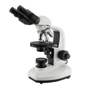
The Trinocular Polarized Microscope uses polarized light to study anisotropic specimens like liquid crystals and minerals. It includes a polarizer positioned in the light path before the specimen and an analyzer placed in the light path between the observation tubes or camera port and objective rear aperture.
The microscope is equipped with two polarizing filters known as polarizer and analyzer. It includes a dividing eyepiece and a trinocular eyepiece tube that is inclined at 30° and can capture the images in 100% light flux. Long infinity objectives are present that make the field of view clear and wide. It also includes 50X ~ 600X magnification lenses, a reflected illumination system, a quadruple nosepiece, a focusing system, a puller-type Bertrand lens as an intermediate attachment, and λ, λ/4, and quarts wedge compensator.
In a polarized microscope, a polarizer transforms white light into plane-polarized light before reaching the sample.
ConductScience offers the Trinocular Polarized Microscope.

Experts in Manufacturing and Exporting Optical Instruments, Microscopes and more other Products.


Specifications | |
|---|---|
| Eyepiece | WF 10X (Φ22mm) Dividing eyepiece (Φ22mm) 0.10mm/Div |
| Eyepiece Tube | Trinocular is inclined 30° and enable to shoot in 100% light flux |
| Objective | Strain-free plan achromatic objective (no cover glass) PL L5X/0.12 (Work distance):26.1 mm PL L10X/0.25 (Work distance):20.2 mm PL L40X/0.60£¨spring£©(Work distance):3.98 mm PL L60X/0.70£¨spring£©(Work distance):3.18 mm |
| Magnification | 50X~600X |
| Reflected Illumination System | 6V30W halogen, brightness enables control. The polarizer can be rotated 360°. The analyzer can be rotatable 360° with scale and minimum vernier Integrated field and aperture diaphragm. |
| Nosepiece | Quadruple (the center of nosepiece is adjustable ) |
| Focus System | Coaxial coarse/fine focus system, with tension adjustable and up stop, minimum division of fine focusing is 2.0μm |
| Intermediate attachment | Puller type Bertrand lens |
| Compensator | λ , λ/4 and quarts wedge compensator |
Documentation
The Trinocular Polarized Microscope uses polarized light to study anisotropic specimens like liquid crystals and minerals. It includes a polarizer positioned in the light path before the specimen and an analyzer placed in the light path between the observation tubes or camera port and objective rear aperture. The light coming from the halogen lamp passes through the polarizer and falls on the birefringent sample that splits the beam; this beam then passes through the analyzer. The final images of the specimen are then captured through the trinocular head camera. The polarizer and analyzer can be rotated from 0°to 360° into or out of the optical path to view the anisotropic sample accurately. Conduct Science’s trinocular polarized microscope comes with a compensator to enhance or reduce the optical path difference.
The microscope is equipped with two polarizing filters known as polarizer and analyzer. It includes a dividing eyepiece and a trinocular eyepiece tube that is inclined at 30° and can capture the images in 100% light flux. Long infinity objectives are present that make the field of view clear and wide. It also includes 50X ~ 600X magnification lenses, reflected illumination system, quadruple nosepiece, focusing system, puller type Bertrand lens as an intermediate attachment, and λ, λ/4, and quarts wedge compensator.
In a polarized microscope, a polarizer transforms white light into plane-polarized light before reaching the sample. The birefringent (double-refracting) specimen splits the beam, which is passed through a second polarizing filter placed parallelly to the polarizer known as an analyzer. Finally, high-contrast images are captured through the trinocular head camera.
The Trinocular Polarized Microscope is known for its applications in the geological sciences, which concentrate on studying minerals in thin rock slices. However, many other materials may conveniently be investigated with polarized light, including natural and manufactured minerals, i.e., ceramics, cement composites, mineral fibers, polymers, urea. The microscope is also used in pharmaceutics, forensic medicines, geology, chemistry, biology, metallurgy, petrology, mineralogy, toxicology, and the discovery of the history of rock formation, starch, wood, and a plethora of biological macromolecules. Some specific applications of the microscope are discussed below:
Monosodium urate crystals are studied using crossed polarization in the polarized microscope.
Collagen is an anisotropic structure that displays the phenomena of birefringence, which the polarized microscope may selectively observe. Collagen fibers serve a crucial function in modeling the biological behavior of different clinical lesions and guiding the epithelium of its bystanders during normal odontogenesis (Kiresur et al., 2019).
Polienko (2019) used the polarized microscope to study uroliths’ surface morphology and mineral composition.
In a study conducted by Pirnstill and Coté (2015), a malarial pigment, hemozoin, was evaluated using a polarized microscope. Polarized light microscopy made it simpler to see the pigment, which was difficult to discern even by experienced laboratory staff.
Synthetic polymer chains tend to organize themselves tangentially after nucleation, and the solidified portions extend outward in a radial fashion. Spherulites, white patches with conspicuous black extinction crossings, are only observed in crossed polarized microscopes.
Keep the microscope away from direct sunlight, extreme humidity, and dust.
The light, lamp housing, and nearby components heat up during usage. Do not handle these pieces until they have cooled. Never try to hold a hot halogen bulb.
Keep the main switch off while changing the halogen lamp, and change the bulb when it has been cooled down
Ensure that the displayed and line voltages are the same. Using different voltages can harm the device.
Don’t adjust the lenses on your own since they already come adjusted and calibrated
The nosepiece and the coarse/fine focus unit have a small and accurate construction, don’t attempt to dismantle them.
The outside surface of the optics should be examined and cleaned frequently using an air stream from an air bulb.
Use the soft cloth or cotton swab dipped in the lens cleaning solution to clean the optical lenses
Clean the lenses in a circular motion, do not use an excessive quantity of solution/solvent
The trinocular polarized microscope is designed to observe anisotropic substances rapidly that are impossible to study with any other microscope. The microscope offers information on absorption color and optical path boundaries between minerals with different refractive indices and differentiates between isotropic and anisotropic materials. This microscope is portable and can be taken anywhere to study the salts in mines. Liquid crystals may be analyzed using the microscope, which is used to look for optical patterns and phase defects. A crystal’s optical positivity or negativity may also be determined using it. It can identify various characteristics of particles, for example, morphology, size, surface texture, hardness, reflectivity, transparency, color, refractive index/indices, magnetism, pleochroism, dispersion staining, birefringence, extinction angle, a sign of elongation, interference figure, fluorescence, and chemical composition.
It uses a very small quantity of samples and can’t study large amounts. It cannot display all the fibers under study. It requires a highly skilled person for its operation.
| Brand | Microscopex |
|---|---|
| eyepiece | |
| objective | PL L10X/0.25 (Work distance):20.2 mm, PL L40X/0.60£¨spring£©(Work distance):3.98 mm, PL L5X/0.12 (Work distance):26.1 mm, PL L60X/0.70£¨spring£©(Work distance):3.18 mm, Strain-free plan achromatic objective (no cover glass) |
| total-magnification |
You must be logged in to post a review.
There are no questions yet. Be the first to ask a question about this product.
Monday – Friday
9 AM – 5 PM EST
DISCLAIMER: ConductScience and affiliate products are NOT designed for human consumption, testing, or clinical utilization. They are designed for pre-clinical utilization only. Customers purchasing apparatus for the purposes of scientific research or veterinary care affirm adherence to applicable regulatory bodies for the country in which their research or care is conducted.
Reviews
There are no reviews yet.