
What are astrocytes?
Astrocytes are a type of glial cells found in the brain and spinal cord. They can be easily distinguishable from other cells because they have a star shape, have numerous branching processes and they represent the most numerous cell populations in the brain, with an estimation that there are 50:1 astrocytes per neuron. There are three types of astrocytes – fibrous, protoplasmatic and radial.
Fibrous astrocytes are found in the white matter and have long unbranched processes. Protoplasmatic astrocytes are found in the grey matter and have short and branched processes. Radial astrocytes can be found between the grey matter and pia mater, in the vertebrate eye and in the cerebellum. Radial astrocytes in the eye make for 23% of the retina and are called Mueller cells. In the cerebellum, they are called Bergmann cells. Astrocytes are connected via gap junctions, meaning that they form an electrically coupled functional connections and this enables them to communicate with neighboring cells as well as with distant ones.
What astrocytes do?
Previously it was considered that astrocytes do not have a specific role other than to fill in the gaps between neurons. Today it is known that astrocytes are involved in a number of brain processes including maintenance of blood-brain barrier, brain homeostasis, nutrition and scarring processes. But besides these functions, astrocytes are involved in neuronal signaling and are able to influence signal propagation. Astrocytes release specific transmitters called gliotransmitters via calcium ion-dependent mechanism and therefore are able to influence neuronal signaling.[1] This phenomenon is called the tripartite synapse consisting of the presynaptic cell, postsynaptic cell, and the glial cell.
We will now discuss the function of astrocytes in greater detail.
- They are the most numerous cells in the brain, surrounding neurons and providing them physical support.
- Astrocytes are providers of nutrients such as lactate. They also provide neurons with glucose during periods of high glucose consumption or glucose depletion.
- Astrocytes help in maintaining the blood-brain barrier and thus play a role in the protection of the brain from pathogens and other harmful substances.
- Given that astrocytes have a role in the maintaining of blood-brain barrier, astrocytes are mediators in neuronal regulation of blood flow into the brain.
- They express neurotransmitter transporters for glutamate, ATP, and GABA and they are also shown to release glutamate and ATP. These characteristics enable them to influence uptake and release of neurotransmitters.
- Since astrocytes have a high number of potassium channels on their outer membrane, when extracellular concentration of potassium rises, astrocytes are able to clear the excessive potassium ions from the extracellular space. This function is very important because high levels of potassium ions in the extracellular space cause cell depolarization and if this happens, uncontrolled firing of neurons would occur.
- Astrocytes help neural repair by forming a glial scar when an injury occurs.
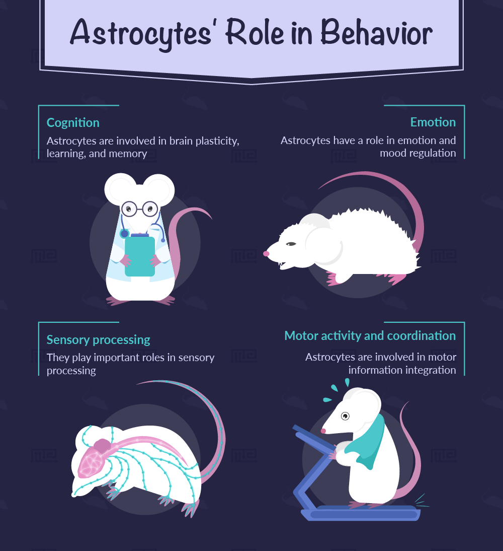
Astrocytes’ role in behavior
As we previously discussed, astrocytes are found around neurons and they can detect neuronal activity because they have neurotransmitter receptors on their membranes. They can release substances such as glutamate, GABA, D-serine, ATP, which can influence the synaptic activity. Following the idea that there is communication between astrocytes and neurons, scientists started examining the possible role astrocytes had in behavior. Since then the evidence for their role in various behaviors has been confirmed. We will now discuss some of the findings.
Cognition
A number of studies showed that astrocytes are involved in brain plasticity, learning, and memory. Tanaka et al. showed that inositol 1,4,5-trisphosphate induced Ca2+ release (IICR) knockout mice exhibited altered behavior in spatial reference memory and remote contextual fear memory.[2] Mice were put in a fear conditioning shock chamber and then were presented with a sound which was followed by a foot shock. The fear learning test was performed after 24h. This part of the test was performed without any auditory cues being presented and their behavior was recorded. After another 24h, the mice were put back in the chamber and tested, but this time with auditory cues. The remote contextual learning test was conducted 30 days after the initial conditioning session. Both knockout mice and controls showed the same level of contextual and cued fear memory after day one and two. But, after 30 days, the knockout mice showed a significantly diminished contextual fear memory when tested without any auditory cues. However, both knockouts and controls showed similar levels of freezing behavior tested for cued fear memory on day 31. This study shows that IICR in the astrocytes is important for remote contextual fear memory.
Although there are a number of studies showing similar results to this experiment, there is still a great necessity to further examine the relation between astrocytes and learning mechanisms.
Emotions
Astrocytes also have a role in emotion and mood regulation. Studies have clearly shown astrocyte involvement in depressive-like behavior in rodents. For example, the study of Banasr and Duman showed that after administration of astrocyte, specific toxin into the prefrontal cortex area of animals showed anhedonia, anxiety-like behavior, and helplessness when tested.[3] In this study, a group of rats was administered with a toxin which ablates astrocytes in the prefrontal cortex while the controls were not. Later the animals were tested and the results showed that the experimental group spent significantly more time immobile in the forced swim test than the controls, which clearly shows signs of helplessness. Additionally, the animals were tested for active avoidance and the experimental group showed increased escape failures compared to controls.
These results show that interfering with the function of astrocytes in the prefrontal cortex leads to depressive-like behaviors in rats and has a clear implication that astrocytes are involved in emotion regulation.
In a study done on connexin 30 knockout mice, Dere et al. found that knockout mice showed higher anxious behavior on open field test compared to controls, but not on rotarod test.[4]
Motor activity and coordination
Although there is diverging evidence about the involvement of astrocytes in general exploratory behavior, some studies showed that in the long term there are some effects.[4] This indicates the need to further explore this topic. However, a study conducted by Paukert et al. suggest motor function engages astrocyte networks in several brain regions via norepinephrine signaling and shows that astrocytes are involved in motor information integration.[5]
Paukert et al.[5] Administered mice with tradzone, a drug that inhibits adrenergic, serotonergic and histaminergic receptors and measured with two photon microscope the Ca2+ transition in Bergmann cells (a type of astrocytes) in the cerebellum. Given that locomotion induces elevation of Ca2+ in Bergmann cells, it was assumed that administration of tradzone would block locomotion induced Ca2+ transition. The behavioral experiment confirmed this assumption. The mice were put on a treadmill with their heads immobilized. The mice could either freely move voluntarily or were forced due to the motor control of the treadmill. The results showed that mice administered with tradzone nearly did not move even in the situation when they were enforced (when the treadmill was controlled by the motor). Tradzone abolished Ca2+ transients in Bergmann cells in both voluntary and enforced locomotion. These results show that norepinephrine activates astrocyte network during locomotion.
Sensory processing
Teubner et al. reported that knockout mice for protein connexin 30, which is expressed in gap junctions of epithelial and mesenchymal tissues in the inner ear, suffer from severe hearing problems.[6] It was discovered that the epithelial tissue starts to degenerate due to the decrease in the potassium levels. The authors of the study indicate that gap junctional signaling is very important for sensory processing and that, given that connexin 30 is also expressed in astrocytes, the deregulation that happens at the gap junction might lead to hearing impairments in the knockout mice.
It was also shown that astrocytes play a role in nociception. Some studies have indicated that astrocytes are involved in the reduction of nociceptive thresholds via gliotransmitters. Foley et al. demonstrated that transgenic mice in which gliotransmission was underexpressed, show lower pain thresholds in the baseline condition than wild type mice when tested in the Von Frey filament test.[7] These animals did not show any pain threshold differences under neuropathic pain. This study suggests that astrocytes mediate pain processing via gliotransmitter regulation.
Tests used to study the role of astrocytes in behavior
Depending on the type of the studied behavior, there are a great number of tests that can show the involvement of astrocytes in behavior.[8] Usually, transgenic animal models or knockout animal models are used for this type of studies. It is also possible to administer drugs to animals so that the desired effect is achieved. But the most essential part of these studies is the behavioral test.
For studying cognition, most of the experiments are exploring spatial working memory in mice. The most common experimental setup includes:
These tests examine exploratory behavior and behavior which leads to escaping aversive stimuli.
When studying emotions and emotional regulation, most of the performed studies were dealing with anxiety-like behaviors as well as depressive-like behaviors.
For testing anxiety-like behavior, researchers commonly use:
These tests measure the amount of exploratory behavior which is diminished in anxious animals.
For testing depressive-like behaviors, the following tests are used:
Their aim is to measure the time the animal will spend actively in a situation with no escape.
When testing motor activity, the researchers use:
- Treadmills
- Rotarod
- Different mazes for assessing exploratory behavior in rodents
In the domain of sensory processing, most of the studies focus on nociception, therefore the following tests are most commonly performed:
- Hotplate test
- Von Frey fiber test
- Foot shock sensitivity test
Clinical relevance of studying the role of astrocytes in behavior
As it was previously shown astrocytes have very important functions in many neuronal processes. Their involvement in the functioning of the central nervous system has been overseen for years, but now a great number of scientists are starting to be aware of their role in the neuronal functioning. Even now, very little is known about the mechanisms astrocytes use to interact not only with neurons and neuronal populations but also among themselves. Having this in mind, one can only assume the lack of knowledge there is about the involvement of astrocytes in neural networks and behavior.
By further examining the role and functioning of astrocytes, there lies a potential in developing new drugs for anxiety and depression. Also, considering that astrocytes are involved in pain processing via gliotransmitters, there is an opportunity to further understand these processes, develop new drugs and treat patients with neuropathic pain. The studies described in the article clearly point to the possibilities for research in various domains of astrocyte functioning and their role in behavior.
References
- Guerra-Gomez, S., Sousa, N., Pinto, L., Oliveira, J.F. (2018), Functional Roles of Astrocyte Calcium Elevations, From Synapses to Behavior, Frontiers in Cellular Neuroscience, vol.11, 427
- Tanaka, M. et al. (2013) Astrocytic Ca2+ signals are required for the functional integrity of tripartite synapses. Mol. Brain 6, 6
- Banasr, M. and Duman, R.S. (2008) Glial loss in the prefrontal cortex is sufficient to induce depressive-like behaviors. Biol. Psychiatry 64, 863–870
- Dere, E. et al. (2003) Connexin30-deficient mice show increased emotionality and decreased rearing activity in the open-field along with neurochemical changes. Eur. J. Neurosci. 18, 629–638
- Paukert, M. et al. (2014) Norepinephrine controls astroglial responsiveness to local circuit activity. Neuron 82, 1263–1270
- Teubner, B. et al. (2003) Connexin 30 (Gjb6)-deficiency causes severe hearing impairment and lack of endocochlear potential. Hum. Mol. Genet. 12, 13–21
- Foley, J.C. et al. (2011) Gliotransmission modulates baseline mechanical nociception. Mol. Pain 7, 93
- Oliveira, J. F., Sardinha, V. M., Guerra-Gomes, S., Araque, A., and Sousa, N. (2015). Do stars govern our actions? Astrocyte involvement in rodent behavior. TrendsNeurosci. 38, 535–549