
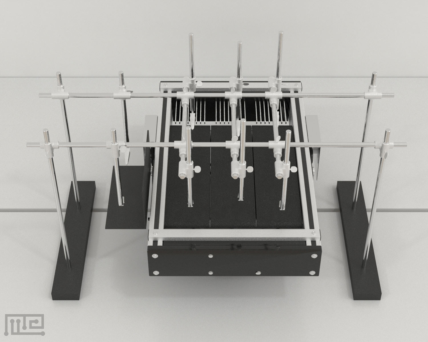
The Treadmill Harness is composed of two parts: harness and body weight support mechanism. The harness is made of a fabric designed as a vest with Velcro straps to fit the animal snuggly. Additional straps are attached to the end of the vest that creates a hook-and-loop mechanism to lift the hind limbs of the animal when attached to the weight support mechanism.
The body weight support mechanism is constructed using adjustable metal clamp systems. Alligator clips attached to the ends of weight support springs are used to attach the harness to the system and support the animal’s weight. The springs can be adjusted to control the weight support the animal receives.
MazeEngineers offers the Treadmill and Treadmill Harness.