
Introduction
The Ancient Greek philosopher Aristotle said that “man is by nature a social animal”. While social behaviors are ubiquitous in the animal kingdom, in no species is sociality more prevalent, more developed and more vital than in human beings. The capacity to be so social, in humans as well as in other mammals, has primarily neurological foundations. This also means that there are many ways in which biological abnormalities can cause a deficit in social abilities.
Given how central social interaction is to human life, and how prevalent disorders affecting social ability are, it is of great importance to understand how social behavior arises from the brain, and how different disorders affect it. The study of both human beings and model organisms such as mice is useful in this endeavor.
In this article, we will start with a brief summary of what social behavior is, why it’s so important and the different kinds of social behaviors shown by humans and other animals. In the second section we’ll discuss the most common human disorders that show a marked impact on social ability. Finally, in the main body of the article, we’ll explore in detail how different parts of the brain have been shown to contribute to skill in social interaction.
Social Behavior: An Overview
Social behaviors are behaviors that involve two or more organisms directly interacting in some way. While these kinds of behaviors are more common in some species than in others, almost all living things engage in a variety of them. Given there are generally a large number of members of a given species alive at a particular point in time, it is almost inevitable that different members will come to interact, and that selection pressures will result in the development of these interactions in an adaptive direction.
Because we rely heavily on social learning, human beings have developed the most extensive culture of any known species, and the richest matrix of social behaviors. We have constructed an extremely interdependent society, reliant on the division of labor and widespread, multi-level cooperation. Hence, being able to engage effectively in social interactions has become a prerequisite for succeeding as a human being, and human beings who suffer from social deficits are at a great disadvantage.
Social behavior is also very important for mice. While mice do not have the same level of culture as humans, they are highly social animals that tend to live in large groups. Common social behaviors between mice include mating, grooming, group sleeping, parenting, genital sniffing, biting, fighting, and squeaking. Most of these behaviors have analogues in humans, who also mate, communicate, parent and engage in aggression. The importance of sociality for mice allows them to serve as models for the research of human social disorders.
Disorders Affecting Social Behavior
In humans, there are many prevalent psychological and physiological disorders that have a detrimental impact on social behaviors. In fact, almost all serious disorders have some kind of impact on social behavior, given how central social interaction is to human life. In this section, we will discuss a few of these disorders. It should be noted that how psychological disorders are officially named and defined is subject to constant revision.
Autism Spectrum Disorders
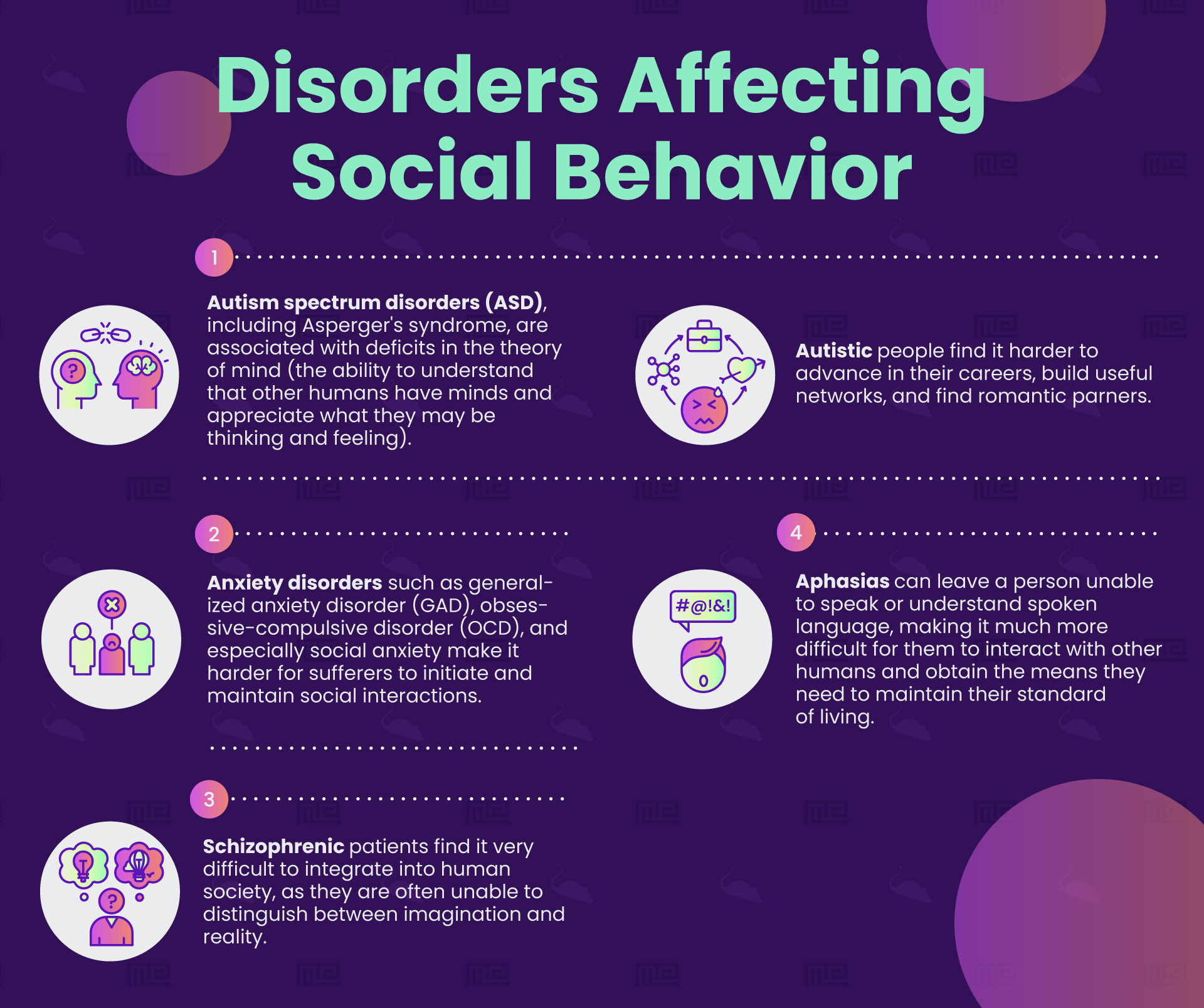
Autism spectrum disorders (ASD), including Asperger’s syndrome, are associated with deficits in theory of mind (the ability to understand that other humans have minds and appreciate what they may be thinking and feeling).[1] These disorders likely have a strong genetic component.[2] The apparent deficit in empathy displayed by autistic individuals, combined with their deficits in understanding and producing language, can lead them to be socially ostracised. Autistic people find it harder to advance in their career, build useful networks and find romantic partners.
Anxiety Disorders
Anxiety disorders such as generalized anxiety disorder (GAD), obsessive compulsive disorder (OCD) and especially social anxiety make it harder for sufferers to initiate and maintain social interactions. Humans with higher than usual anxiety in social situations may avoid such situations, slowing down the development of their social skills, and making it harder for them to form useful and meaningful relationships. The development of anxiety disorders is strongly associated with both genetic and environmental factors.[3]
Schizophrenia
Schizophrenia denotes a loosely defined cluster of disorders that all exhibit an apparent detachment from reality as a major symptom. It is thought by some to be a condition unique to humans.[4] Schizophrenic patients find it very difficult to integrate into human society, as they are often unable to distinguish between imagination and reality. Moreover, paranoid schizophrenics may refuse to form relationships with others on the basis of distrust. Schizophrenia has been strongly linked with genetic factors.[5]
Language Disorders
Disorders of language, known as aphasias, come in various kinds and all have an impact on social behavior. A key component for humans in engaging in complex social behaviors is the ability to produce and understand language. Aphasias, often caused by stroke or brain damage, can leave a person unable to speak or understand spoken language, making it much more difficult for them to interact with other humans and obtain the means they need to maintain their standard of living.
Non-Neurological Disorders
While social deficits are more commonly associated with psychological disorders such as those detailed above, non-psychological issues can also impact social behavior. For example, someone who has difficulty walking due to a spinal cord injury will be less able to attend social events and so will engage in fewer useful social interactions. If mobility is restricted from a young age, the development of basic social skills may be severely and permanently affected.[6]
The Neuroscience of Social Behavior
An entire subfield of neuroscience, known as social neuroscience, is dedicated to understand how the brain produces and deals with social behaviors. As with all major behavioral areas, there is no one single part of the brain that is dedicated to social interaction, but rather a number of different brain regions that contribute to different aspects of the behaviors.
The Mirror Neuron System
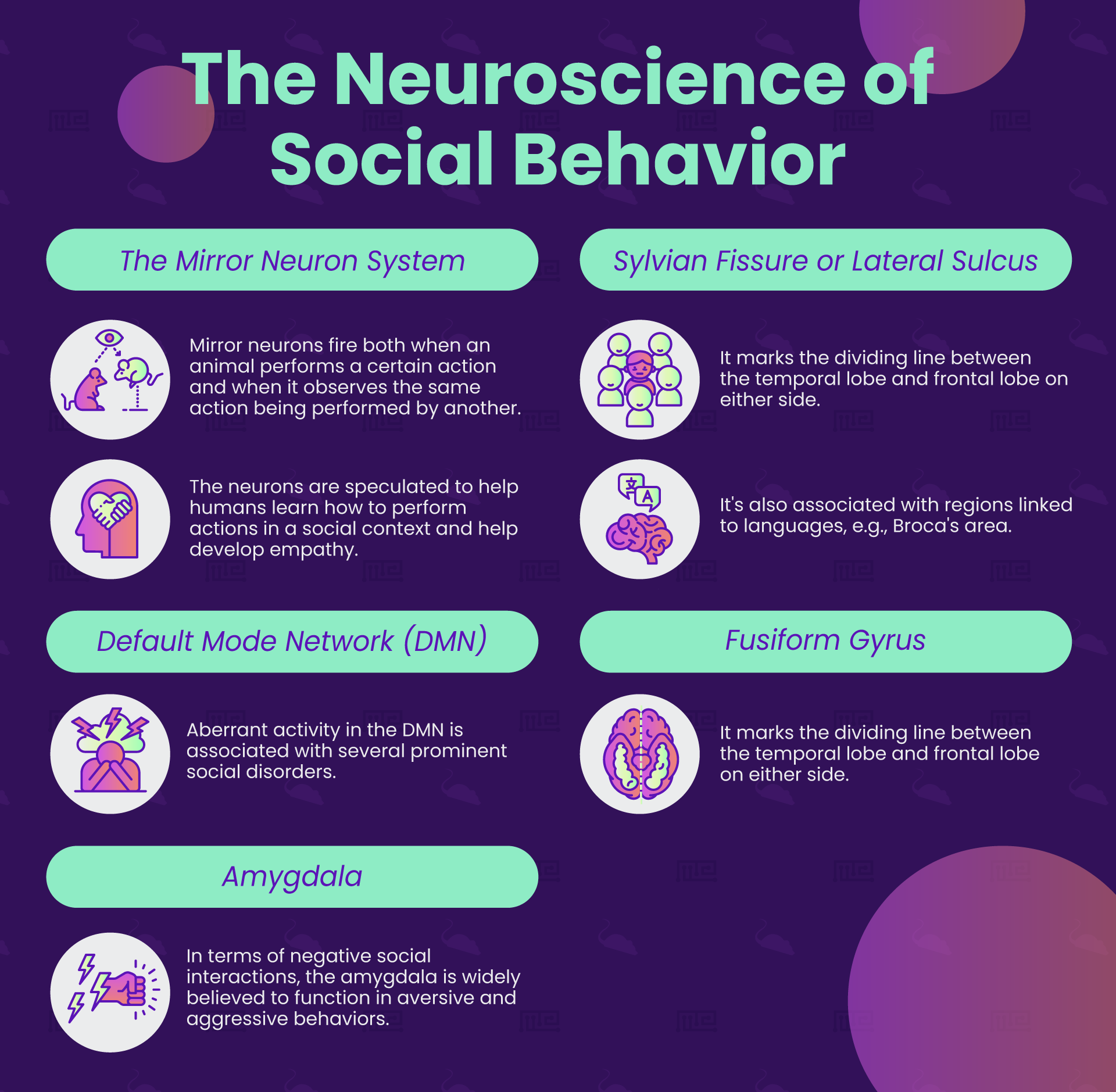
Mirror neurons were first discovered in the brains of macaques;[7] there is evidence they are also present in the brains of humans and rats, as well as other creatures.[8][9] Mirror neurons are neurons that fire both when an animal performs a certain action, and when that animal observes the same action being performed by another individual. Hence, the neurons directly “mirror” the activity of another living system. Much speculation (often ungrounded) has been made about mirror neurons since their discovery, and it is important not to stray too far beyond what is scientifically known.
Imitating Behavior
Social learning is extremely important for human beings, perhaps more so than for any other species. Humans engage in many complicated daily activities—driving a car, using kitchen appliances, operating a computer and so on—that cannot be encoded in genetic instructions, and have to be learned through imitation. Humans appear to have a compulsion for social imitation, exemplified by children’s desire to copy whatever they see, and by adults doing things such as adopting accents and mannerisms. It could be that stereotyped practices are literally contagious, spreading through the human population via emulation in mirror neurons.
Mirror System and the Motor System
Neurons whose behavior resembles macaque mirror neurons are thought to exist primarily in the parts of the human brain associated with movement, such as the somatosensory cortex, premotor cortex and supplementary motor area.[10] It has been speculated not only that these neurons help humans learn how to perform actions in a social context, but also that the way they simulate what is happening inside another individual’s nervous system allows us to develop an understanding of how it feels to be another individual, and so supports theory of mind.
fMRI Findings
We must be aware however that none of these predictions have so far been demonstrated. While fMRI studies suggest mirror neurons are present in humans, this has not been confirmed. It has not been explained in detail how the computations carried out by mirror neurons could contribute to mimesis or theory of mind. Also, if mirror neurons are indeed a mechanism for social imitation, it is strange that macaques possess them since they do not seem to rely on social learning much at all. Thus, a lot more research must be conducted before any definitive statements about a human mirror neuron system can be made.
Default Mode Network
The default mode network (DMN) is a large network of regions in the brain that tend to be active together. It includes the hippocampus, parahippocampus, posterior cingulate cortex, medial prefrontal cortex, and parts of the parietal and temporal cortex. It is known as the default mode network because it was first characterized as being a network that tends to activate when a person is at rest, engaging in passive activities like mind-wandering and daydreaming. However, more recent research has associated this network with social behaviors.
Aberrant activity in the DMN is associated with several prominent social disorders. For example, individuals with attachment issues as a result of parental abuse during childhood show lower connectivity in the DMN, as do people with post-traumatic stress disorder.[11] Lower DMN connectivity is also seen with autism spectrum disorders, and appears to positively correlate with the severity of the autism.[12] At the other extreme, greater than usual DMN connectivity has been linked with rumination, the tendency to become pathologically fixated with one’s own psychological symptoms in a way that prevents treating them.[13]
Fusiform Gyrus
The fusiform gyrus is a region at the back of the brain near the cerebellum whose full function is not entirely understood, but that is most commonly linked with recognizing faces. Individuals with lesions to the fusiform gyrus can suffer from the disorder prosopagnosia, or “face blindness”, and are not able to recognize people by their faces.
Since humans build and rely on uniquely complex social networks, as well as leaning a lot on our sense of sight to navigate the world, it is especially important for humans to be able to remember and promptly recognize a large number of other individuals. Similar mechanisms specialized for processing facial data appear to be present in macaques and chimpanzees as well, although fMRI scans show activity is not concentrated in the same fusiform gyral region in these other primates.[14] Mice, who rely much more on scent to recognize others, are not believed to have similar facial recognition mechanisms.
Facial Recognition
Faces occupy an extremely important position in human social life. The human brain is so well-honed to recognize faces that it even hallucinates them, leading humans to become enthralled by and even impute sentience to inanimate objects with “faces” on them. However, one group that diverges sharply from this trend is patients with autism spectrum disorders. Autistic individuals show a lower density of neurons in the fusiform face area.[15] This is associated with greater difficulty in recognizing faces, and a general lack of interest in faces compared to inanimate objects.
Amygdala
The amygdala, a small almond-shaped region in the temporal lobes of the brain, is popularly associated with fear and the “fight or flight” response. In truth, the amygdala seems to play a role in a broad range of behaviors, including both positive and negative social interactions.
Function
In terms of negative social interactions, the amygdala is widely believed to play a function in aversive and aggressive behaviors. Stimulation of the amygdala encourages more aggressive and sexual behavior, whereas damage to the amygdala results in passivity and the absence of normal fear responses.[16] fMRI studies also show that the amygdala is active when an individual perceives their personal space to be violated.[17] It thus seems the amygdala is helping with defensive or dominance functions.
Amygdala Cell Death
In mice, a 2018 study carried out by a research team in the United States showed that individuals possessing a mutant allele of the transcription factor Tshz1 showed a marked loss of regulatory cells in the hippocampus. This cell death was associated with a number of deficits, such as increased fear and depression and passivity in social situations. In a direct social interaction test, mutant mice spent 95% less time than controls actively engaging in social interaction, and also spent much more time performing non-social tasks such as self-grooming and solitary exploration. Mutant mice seemed uninterested in establishing a position for themselves in the social hierarchy, and were much more vulnerable to aggression from other mice.[18]
Amygdala Deficits
Another mouse study, this time from 2015, found correlates of social deficits in the amygdala of the mouse strain BTBR T+Itpr3tf/J, which is used in the study of autism. Experimental animals undertook the social proximity test and fear conditioning. BTBR T+Itpr3tf/J mice showed less physical contact than control (C57BL/6J) mice, and showed an unusual avoidant response to a stressed cagemate. These behavioral abnormalities were correlated with reduced c-Fos expression in the amygdala, hippocampus and prefrontal cortex of BTBR T+Itpr3tf/J compared to controls.[19]
The volume of the amygdala in humans has been positively correlated with a number of positive social factors. Those with larger amygdalae on average have more extensive and more densely interconnected social networks, as well as an improved ability to judge the emotions of others via their facial expressions.[20][21] It has been hypothesized that the amygdala plays a central part in emotional intelligence, and so humans with larger amygdalae have greater emotional intelligence leading to more success in forming and maintaining relationships.
Sylvian Fissure
The Sylvian fissure, also known as the lateral sulcus, is a distinctive feature of the brain that marks the dividing line between the temporal lobe and frontal lobe on either side. It is in the Sylvian fissure on the left side of the brain where regions long associated with language, such as Broca’s area and Wernicke’s area, are found.
Symbolic Level of Cognition
No other species uses and relies on language to the same extent as humans. The neuroscientist Terrence Deacon has argued that humans possess a unique “symbolic” level of cognition which allows us to understand and express abstract concepts. This, in turn, allows us to plan far into the future, form contracts and develop other complex social relations.[22] While humans without language could still communicate in some ways, such as through gestures or calls, they would be extremely limited in their ability to participate in all the most common social behaviors.
Aphasia
The term aphasia, as mentioned earlier, describes disorders of language in humans. There are many different kinds of aphasia corresponding to different aspects of language function; these include issues in articulating words, understanding words, organizing words grammatically and using the right words in the right situation. These can be caused by damage to the brain, or by a failure to acquire language during the crucial window in childhood when the brain is most receptive to language learning.
Broca’s Aphasia
Damage to Broca’s area leads to Broca’s aphasia, where a patient has difficulty producing language. They can understand what is said to them, but tend to stutter and miss out conjunctives and other functional words when trying to form sentences. Broca’s aphasia also affects sign language ability in deaf patients, suggesting that Broca’s area is not merely involved in the bare motor articulation of sounds, but rather in the more abstract function of generating language per se.
Wernicke’s Aphasia
Damage to Wernicke’s area causes Wernicke’s aphasia, where a patient produces fluent but meaningless verbiage, has trouble understanding what is said to them and also finds it harder to retrieve target words. They may invent entirely new words or alter existing words. Both Broca’s aphasia and Wernicke’s aphasia are commonly caused by either brain hemorrhage or traumatic brain injury. It should be noted that patients are not always easily sorted into one aphasia or the other, since brain damage rarely affects just one functional region.
Conclusion
Over the past few decades, neuroscience has made great strides in understanding how the brain enables social behaviors, and how various disorders affecting the nervous system can inhibit an individual’s ability to engage in social interaction. While there is still a long way to go until the neuroscience of social behavior is fully understood, the insights already uncovered assist the development of medical treatments, and provide a platform for deeper investigation.
References
- Brewer, N., Young, R. L., & Barnett, E. 2017. Measuring Theory of Mind in Adults with Autism Spectrum Disorder. Journal of autism and developmental disorders, 47(7), 1927–1941.
- Yoo H. 2015. Genetics of Autism Spectrum Disorder: Current Status and Possible Clinical Applications. Experimental neurobiology. 24(4), 257–272.
- ScienceDaily. 2019. Social anxiety is highly heritable but is affected by environment — ScienceDaily. [ONLINE] Available at: https://www.sciencedaily.com/releases/2016/01/160120092655.htm. [Accessed 26 October 2019].
- Bret Stetka. 2019. Why Don’t Animals Get Schizophrenia (and How Come We Do)? – Scientific American. [ONLINE] Available at: https://www.scientificamerican.com/article/why-don-t-animals-get-schizophrenia-and-how-come-we-do/. [Accessed 26 October 2019].
- Tyrone D. Cannon; Jaakko Kaprio; Jouko Lönnqvist; Matti Huttunen; Markku Koskenvuo. 1998. The genetic epidemiology of schizophrenia in a Finnish twin cohort A population-based modeling study. Archives of General Psychiatry. 55 (1): 67–74.
- Marzi, I., & Reimers, A. K. 2018. Children’s Independent Mobility: Current Knowledge, Future Directions, and Public Health Implications. International journal of environmental research and public health. 15(11), 2441.
- Di Pellegrino, G.; Fadiga, L.; Fogassi, L.; Gallese, V.; Rizzolatti, G. 1992. Understanding motor events: a neurophysiological study. Experimental Brain Research. 91 (1): 176–180.
- Gazzola, V.; Keysers, C. 2009. The observation and execution of actions share motor and somatosensory voxels in all tested subjects: single-subject analyses of unsmoothed fMRI data. Cereb Cortex. 19 (6): 1239–1255.
- Carrillo, M., Han, Y., Migliorati, F., Liu, M., Gazzola, V., & Keysers, C. 2019. Emotional Mirror Neurons in the Rat’s Anterior Cingulate Cortex. Current biology : CB, 29(8), 1301–1312.
- Keysers, Christian; Gazzola, Valeria. 2010. Social Neuroscience: Mirror Neurons recorded in Humans. Current Biology. 20 (8): R353–354.
- Akiki, Teddy J.; Averill, Christopher L.; Wrocklage, Kristen M.; Scott, J. Cobb; Averill, Lynnette A.; Schweinsburg, Brian; Alexander-Bloch, Aaron; Martini, Brenda; Southwick, Steven M.; Krystal, John H.; Abdallah, Chadi G. 2018. Default mode network abnormalities in posttraumatic stress disorder: A novel network-restricted topology approach. NeuroImage. 176: 489–498.
- Washington, Stuart D.; Gordon, Evan M.; Brar, Jasmit; Warburton, Samantha; Sawyer, Alice T.; Wolfe, Amanda; Mease-Ference, Erin R.; Girton, Laura; Hailu, Ayichew. 2014. Dysmaturation of the default mode network in autism. Human Brain Mapping. 35 (4): 1284–1296.
- Zhu, X; Wang, X; Xiao, J; Liao, J; Zhong, M; Wang, W; Yao, S. 2012. Evidence of a dissociation pattern in resting-state default mode network connectivity in first-episode, treatment-naive major depression patients. Biological Psychiatry. 71 (7): 611–7.
- Yovel, G., & Freiwald, W. A. 2013. Face recognition systems in monkey and human: are they the same thing?. F1000prime reports, 5, 10.
- Van Kooten IA, Palmen SJ, von Cappeln P, Steinbusch HW, Korr H, Heinsen H, Hof PR, van Engeland H, Schmitz C. 2008. Neurons in the Fusiform Gyrus are Fewer and Smaller in Autism. Brain. 131 (4): 987–99.
- ScienceDaily. 2019. Aggression. [ONLINE] Available at: https://www.sciencedaily.com/terms/aggression.htm. [Accessed 26 October 2019].
- Kennedy DP, Gläscher J, Tyszka JM, Adolphs R (October 2009). “Personal space regulation by the human amygdala”. Nature Neuroscience. 12 (10): 1226–7.
- Jeffrey Kuerbitz, Melinda Arnett, Sarah Ehrman, Michael T. Williams, Charles V. Vorhees, Simon E. Fisher, Alistair N. Garratt, Louis J. Muglia, Ronald R. Waclaw and Kenneth Campbell. 2018. Journal of Neuroscience. 38 (5). 1160-1177.
- Meyza K, Nikolaev T, Kondrakiewicz K, Blanchard DC, Blanchard RJ and Knapska E. 2015. Neuronal correlates of asocial behavior in a BTBR T+Itpr3tf/J mouse model of autism. Front. Behav. Neurosci. 9:199.
- Bickart KC, Wright CI, Dautoff RJ, Dickerson BC, Barrett LF. 2011. Amygdala volume and social network size in humans. Nature Neuroscience. 14 (2): 163–4.
- Bzdok D, Langner R, Caspers S, Kurth F, Habel U, Zilles K, Laird A, Eickhoff SB. 2011. “ALE meta-analysis on facial judgments of trustworthiness and attractiveness”. Brain Structure & Function. 215 (3–4): 209–23.
- Deacon, Terrence. 1997. The Symbolic Species. W.W. Norton & Co.