
Usually, in behavioral and brain sciences, the neurons play the lead character and get most of the attention. After all, neurons are the brain’s basic cell for transmitting signals to other neurons or muscles.
However, glial cells, which play the supporting role, are also vital for behavior. In this article, we will take a closer look at how microglia, a particular type of glial cell, are involved in behavior.
What Are Glial Cells?
As mentioned previously, glial cells are like supporting characters of the brain. Essentially, they help and maintain neuronal integrity.
There are many different types of glial cells.
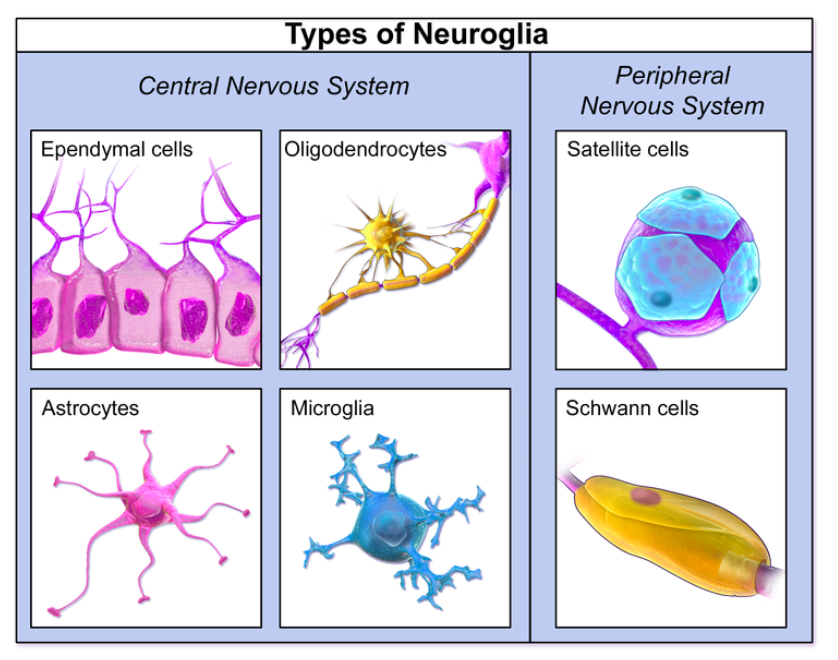
In the central nervous system (CNS), the following glial cells can be found: astrocytes, oligodendrocytes, ependymal cells, and microglia.
The peripheral nervous system contains the following glial cells: satellite cells and Schwann cells.
Glial cells have five main functions:
- to surround neurons and hold them in place.
- to supply nutrients and oxygen to neurons.
- to insulate one neuron from another.
- to destroy pathogens and remove dead neurons.
- sometimes, to aid in neurotransmission and synaptic connections.
What Are Microglia?
Microglial are dynamic, plastic cells that can adapt to specific CNS conditions in order to carry out a tailored immune response.
Traditionally, microglia were seen solely as immune cells responsible for fighting bacteria or other exogenous substances which found their way into the brain.
However, microglial cells are ultimately able to affect behavior and their integrity is necessary for optimum functioning.
Although understanding how microglia communicate with other parts of the CNS is a complicated process, research is increasingly showing that neurons and microglia interact.
For example, interactions between microglia and hippocampal neurons have many implications when it comes to learning and memory, two key cognitive processes driving behavior.[1]
Function of Microglia
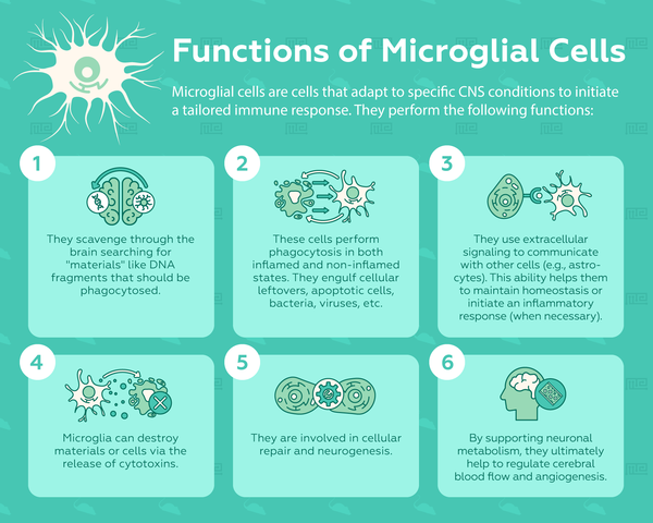
Even though microglia make up <10% of the brain’s total cells,[2] they have many important roles to fill. Below is an overview of microglial functions:
- Perform Phagocytosis: Phagocytosis is the uptake or ‘eating’ of materials. Microglia cells commonly engulf lipids, cellular leftovers, apoptotic cells (when in the non-inflamed state), and foreign materials like viruses or bacteria (in the inflamed state).
- Able to Scavenge: Microglial cells can regularly scavenge and move through brain regions, ultimately acting as a form of housekeeping. If any: plaques, DNA fragments, apoptotic cells, foreign materials, damaged cells, or neurofibrillary tangles are found, then microglial phagocytosis will be activated. And, microglia will ‘eat’ these materials in order to get rid of them.
- Communicate via Extracellular Signaling: Microglia communicate with other cells (and astrocytes, nerves, etc.) via a complex chain of extracellular signaling molecules. This function is important for two reasons: to maintain homeostasis in a non-inflamed state or to promote an inflammatory response in damaged or infected tissue in order to resolve the issue and return to homeostasis.
- Release cytotoxic substances: By releasing cytotoxic substances, microglia can destroy other materials or cells, a process that supplements phagocytosis.
- Cellular repair promoters: Microglia have been associated with cellular repair and also the modulation of neurogenesis.
- Support neuronal metabolism: Supporting neuronal metabolism is another microglial function that is important for homeostasis as it helps regulate cerebral blood flow and angiogenesis.[3]
Since microglia have so many important and complex functions, they are indispensable throughout all stages of development.
However, much about microglial function remains unknown. Thus, researchers are trying to get a better understanding of how microglial function is related to behavior by manipulating microglia and studying subsequent outcomes. Check out our article on Microglial Physiology and Behavior and see how behavioral researchers are using knockout mice, for example, to uncover the relationship between microglial physiology and behavior.
Factors Leading to Microglial Dysfunction
However, some factors alter the microglial cells’ efficiency and make them dysfunctional, including:[4]
- Aging: Aging is associated with the loss of cell function. Check out our supplemental article on Microglia, Aging, and Behavior to see what studies have shown how behavior is influenced by aging and microglia.
- Genetics: Genetic risk factors may influence microglial integrity.
- Epigenetics: For example, histone modifications can influence gene expression, ultimately affecting cellular plasticity or the inflammatory response.[4][5]
Microglia: The CNS’s immune cells
Microglia are the guardians of the CNS. They are the main immune response for any damage injury, pathology, or infection relating to the brain and spinal cord.
Microglia are often compared to macrophages (a large white blood cell that can perform phagocytosis and eat other cells).
The difference between microglia and macrophages, however, is that microglia are able to transform without causing an immunological imbalance.[6]
Microglia are very plastic and can change structurally depending on location and the precise needs at that point in time. Thus, microglial form varies according to the detected chemical signals and relevant conditions.
Microglial Phenotypes
Depending on what information microglia receive from their environment, they can have different phenotypes:
- M1 Phenotype: The classical activation phenotype which occurs in response to bacterial or viral infection.
- M2 Phenotype: The alternative or anti-inflammatory phenotype which occurs in neurodegenerative diseases or when neuronal cell death is detected.
However, this M1/M2 polarization paradigm has been increasingly criticized and questioned as possibly being overly simplistic since it is now recognized that microglial activation occurs on a continuum.[5]
Furthermore, a third phenotype has also emerged recently, one where microglia are pro-tumoral and able to support tumor growth.[7]
These phenotypes demonstrate the versatility and complexity of microglial cells.
Inflammation has been implicated in behavioral change via microglial activity. Thus, this is a particular area of interest for researchers studying the interplay between these factors. We cover this area in-depth (while providing research examples) in the article Microglia, Inflammation, and Behavior.
Microglia and Disease
Microglia are crucial for keeping the brain healthy and in homeostasis. But, when microglia are over-activated or uncontrolled, they are implicated in brain-related diseases.
Overactive microglia have been linked to neurodegenerative diseases such as:
- Alzheimer’s disease
- Parkinson’s disease
- Huntington’s disease
- Amyotrophic lateral sclerosis[5]
While uncontrolled microglia have been associated with increased levels of tumor cell migration.[5]
Inflammation is also very likely to be implicated in these diseases somehow, too.
Ultimately, these diseases have an impact on behavior and, at the same time, may cause/be influenced by altered microglial levels. Thus, to learn more about how behavioral scientists are exploring this area of research, check out our article Microglia, Diseases, and Behavior.
How Do You Connect Microglia and Behavior Together?
It’s not an easy task to tie microglia with behavior as there are many factors involved in cell signaling and communication.
In order to establish that microglia have influenced behavior and are involved in the chain of events leading up to behavior, many complex techniques and assessments are needed.
Microglial activation is measured through the help of biomarkers and histological techniques. Scientists can also use PET scans, dissect the brain and stain slices, and immunochemistry, for example, as ways to quantify and compare microglial abundancy and activation across various experimental conditions.
Cytokines can also be measured as a proxy of microglial activation. Also, measurements of CD11b (also known as C3R) and Iba1 are used as microglial biomarkers.[8][10]
Microglial Cytokines and Markers
In order to measure microglial activity and expression, experiments quantify microglial cytokines and markers.
Microglia can release cytokines (small proteins crucial for cellular signaling). The following are cited frequently in scientific literature as cytokines indicating microglial activation:
The following microglia markers are also measured as a means of quantifying microglial activity:
In order to be able to implicate microglia in behavioral changes, there needs to be a significant alteration in microglial markers and cytokines.
Conclusion
Although the intersection of microglia and behavior is a relatively new area of interest for researchers, it is rapidly growing due to its promising application for resolving health-related issues or developing pharmaceutical solutions for disease.
Since the relationship between behavior and microglia has been established, future directions will focus on uncovering the mechanisms that make this possible, including the role that other systems play like microbiota.[13][14]
Also, future directions will begin to study the combined effects of environmental stressors on microglial activity and behavior. For more information on the relationship between the CNS immune cells and the environment, check out our article on Environmental Effects on Microglia and Behavior.
References
- Wake, Hiroaki, Andrew J. Moorhouse, and Junichi Nabekura. “Functions of microglia in the central nervous system–beyond the immune response.” Neuron glia biology 7.1 (2011): 47-53.
- Lawson, Linda J., et al. “Heterogeneity in the distribution and morphology of microglia in the normal adult mouse brain.” Neuroscience 39.1 (1990): 151-170.
- Blank, Thomas, and Marco Prinz. “Microglia as modulators of cognition and neuropsychiatric disorders.” Glia 61.1 (2013): 62-70.
- Streit, Wolfgang J. “Microglia as neuroprotective, immunocompetent cells of the CNS.” Glia 40.2 (2002): 133-139.
- Cheray, Mathilde, and Bertrand Joseph. “Epigenetics control microglia plasticity.” Frontiers in cellular neuroscience 12 (2018): 243.
- Gehrmann, Jochen, Yoh Matsumoto, and Georg W. Kreutzberg. “Microglia: intrinsic immuneffector cell of the brain.” Brain Research Reviews 20.3 (1995): 269-287.
- Hambardzumyan, Dolores, David H. Gutmann, and Helmut Kettenmann. “The role of microglia and macrophages in glioma maintenance and progression.” Nature neuroscience 19.1 (2016): 20.
- Kopec, Ashley M., et al. “Microglial dopamine receptor elimination defines sex-specific nucleus accumbens development and social behavior in adolescent rats.” Nature communications 9.1 (2018): 3769.
- Jeong, Hey-Kyeong, et al. “Brain inflammation and microglia: facts and misconceptions.” Experimental neurobiology 22.2 (2013): 59-67.
- Wang, Hong-Tao, et al. “Early-life social isolation-induced depressive-like behavior in rats results in microglial activation and neuronal histone methylation that are mitigated by minocycline.” Neurotoxicity research 31.4 (2017): 505-520.
- Galic, Michael A., Kiarash Riazi, and Quentin J. Pittman. “Cytokines and brain excitability.” Frontiers in neuroendocrinology 33.1 (2012): 116-125.
- Smith, Joshua A., et al. “Role of pro-inflammatory cytokines released from microglia in neurodegenerative diseases.” Brain research bulletin 87.1 (2012): 10-20.
- Erny, Daniel, et al. “Host microbiota constantly control maturation and function of microglia in the CNS.” Nature neuroscience 18.7 (2015): 965.
- 1Shemer, Anat, et al. “Microglia plasticity during health and disease: an immunological perspective.” Trends in immunology 36.10 (2015): 614-624.