
Microbiology
Basic Microbiology Techniques
Are you looking for Laboratory Devices for your lab? Click here Overview There are several microbiology techniques and procedures specially developed over the years to


Are you looking for Laboratory Devices for your lab? Click here Overview There are several microbiology techniques and procedures specially developed over the years to
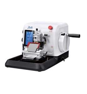
Our AccuSection Rotary microtome is an automatic, semi-automatic, and manual Microtome that meets the different needs of microtomists that cuts the sections of biological specimens into thin slices for use in microscopy. It is used to prepare thin layers of bone, minerals, teeth, and hair with section thicknesses ranging from 1 micron to 60 microns. Hard materials that use synthetic resins, they can slice up to 0.5 microns.
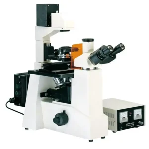
An inverted fluorescent microscope uses fluorescence to study specimens, such as living cells at the bottom of a petri dish or tissue culture. The Inverted Fluorescent Microscope has similar components to those of other microscopes, and the only difference is the arrangement of the parts, which are placed in inverted positions.
The specimen is lit using a halogen lamp as a light source. When light enters the microscope, it strikes a dichroic mirror, reflecting one range of wavelengths while allowing another to pass through. The ultraviolet light is reflected up to the specimen by the dichroic mirror. The UV light causes fluorescence in the specimen’s molecules and the fluorescent-wavelength light generated is collected by the objective lens.
ConductScience offers the Inverted Fluorescent Microscope.
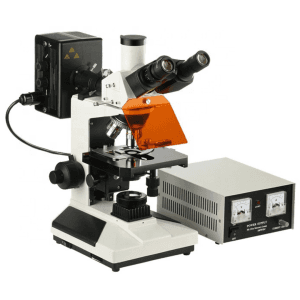
The Trinocular Mercury Lamp Fluorescence Microscope utilizes the principle of fluorescence to illuminate the object by using a mercury lamp as a light source. The mercury lamp gives accurate, fast, and reliable results due to its ability to produce bright spectral lines within the visible region of the UV spectrum.
The Trinocular Mercury Lamp Fluorescence Microscope is equipped with two eyepieces (binocular) and an additional third eyepiece tube to connect a microscope camera, making it trinocular. It has a focus system consisting of fine and coarse adjustment knobs. It also contains a quadruple-reversed nosepiece, double-layer mechanical working stage, abbe condenser, collector for placing a halogen lamp, and blue and frosted filters. The light source is provided by a mercury lamp and a halogen lamp.
ConductScience offers the Trinocular Mercury Lamp Fluorescence Microscope.

The Halogen Lamp Fluorescence Microscope produces images of stained tissues, animal and microbiological cell colonies, and pathogens irradiated with light. It utilizes specific wavelengths of light, particularly infrared light, to excite fluorophores in specimens infused with the fluorochrome pigment and thereby generates a highly illuminated image with distinguishable boundaries.
The primary purpose of the halogen lamp fluorescence microscope is to irradiate the specimen with a specific wavelength of light, separate the weaker emitted light from the bright excitation light, and only allow the emitted light to reach the detector. When light reaches the detector, the fluorescent structure of the specimen is superimposed against a deep, dark, almost black background.
ConductScience offers the Halogen Lamp Fluorescent Microscope.

The Trinocular Polarized Microscope uses polarized light to study anisotropic specimens like liquid crystals and minerals. It includes a polarizer positioned in the light path before the specimen and an analyzer placed in the light path between the observation tubes or camera port and objective rear aperture.
The microscope is equipped with two polarizing filters known as polarizer and analyzer. It includes a dividing eyepiece and a trinocular eyepiece tube that is inclined at 30° and can capture the images in 100% light flux. Long infinity objectives are present that make the field of view clear and wide. It also includes 50X ~ 600X magnification lenses, a reflected illumination system, a quadruple nosepiece, a focusing system, a puller-type Bertrand lens as an intermediate attachment, and λ, λ/4, and quarts wedge compensator.
In a polarized microscope, a polarizer transforms white light into plane-polarized light before reaching the sample.
ConductScience offers the Trinocular Polarized Microscope.
The Series Polarizing Microscope with Halogen Lamp uses reflected and transmitted light to observe birefringent materials such as rocks and polymers. The halogen lamp is used as a light source, and a pair of polarizing filters produce polarized light.
The microscope is equipped with an eyepiece, eyepiece tube, blue and frosted filters, polarizer, and collector. It supports both binocular and trinocular heads that can be inclined at 30°. It comes with 4 achromatic objective lenses with different magnifications. In addition, it has a quadruple nosepiece sliding polarizing analyzer, 120 mm rotatable, and double layer mechanical stage with 75mm×35mm moving range which helps not only in moving the stage in a vertical direction but also along the horizontal axes. The coaxial coarse and fine focus system includes a minimum division of fine focusing up to 2 microns. An Abbe condenser is used to focus the light onto the specimen. Furthermore, a 6V 20W halogen lamp and a polarizer are used to illuminate the sample.
ConductScience offers the Polarizing Microscope with Halogen Lamp.

The trinocular inverted microscope is a high-level microscope designed for research institutes, health and medical units, and universities to observe cultured living cells.
The trinocular inverted microscope comes with 2 eyepieces and a third eye tube for a better viewing experience. The third eye tube allows the microscope to be connected to a camera for image capturing and video recording, which adds to its versatility in using varying laboratory studies. It can be used to study live-cell imaging of cancer cells, the combined effect of EMF and low-level lenses on biological processes, and study the chitin-based scaffolds, etc.
ConductScience offers the Trinocular Inverted Microscope

The Digital Inverted Microscope is equipped with advanced video and image processing and is best for precise laboratory cell analysis and live-cell imaging and recording.
The Digital Inverted Microscope comes with a total magnification between 100X and 400X. It is connected with a display screen or microscope camera to view the image of the specimen under observation. Also, it has a double-layer mechanical stage (with horizontal and vertical adjustable knobs) and three-sized petri dish trays, allowing you to observe different samples. With an inbuilt 2.0 USB interface, the microscope can transfer data at high speed.
ConductScience offers the Digital Inverted Microscope