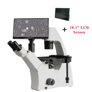
Scientists Record the Assembly of Ribosome in Real-Time
Scientists develop a new method for monitoring ribosome formation in real-time. The new perspective is changing our understanding of cell biology and inner-cellular processes.
Resources available for scientists, science students, and researchers.
Scientists develop a new method for monitoring ribosome formation in real-time. The new perspective is changing our understanding of cell biology and inner-cellular processes.
The Trinocular Mercury Lamp Fluorescence Microscope utilizes the principle of fluorescence to illuminate the object by using a mercury lamp as a light source. The mercury lamp gives accurate, fast, and reliable results due to its ability to produce bright spectral lines within the visible region of the UV spectrum.
The Trinocular Mercury Lamp Fluorescence Microscope is equipped with two eyepieces (binocular) and an additional third eyepiece tube to connect a microscope camera, making it trinocular. It has a focus system consisting of fine and coarse adjustment knobs. It also contains a quadruple-reversed nosepiece, double-layer mechanical working stage, abbe condenser, collector for placing a halogen lamp, and blue and frosted filters. The light source is provided by a mercury lamp and a halogen lamp.
ConductScience offers the Trinocular Mercury Lamp Fluorescence Microscope.
As an Amazon Associate, ConductScience Inc. earns revenue from qualifying purchases Introduction Fungi is a group of eukaryotic organisms that include a myriad of single and
Reference to this article: ConductScience, Estimating the Chiasma Frequency in Diplotene-Diakinesis Stage (2019). doi.org/10.55157/CS20200925 Introduction Chiasma is the point of crossing over or site where the
The information about the structure, interactions, and dynamics of biomolecules (protein, DNA, and RNA) is essential for understanding cellular processes. This knowledge is deconvolved by scientists
Need Microscopes for your Lab? Click here Stereo Microscopes: Introduction Microscopes are sophisticated instruments used to see and analyze small specimens. With impressive optical and mechanical
Are you looking for Laboratory Devices for your lab? Click here Overview There are several microbiology techniques and procedures specially developed over the years to study
The Trinocular Polarized Microscope uses polarized light to study anisotropic specimens like liquid crystals and minerals. It includes a polarizer positioned in the light path before the specimen and an analyzer placed in the light path between the observation tubes or camera port and objective rear aperture.
The microscope is equipped with two polarizing filters known as polarizer and analyzer. It includes a dividing eyepiece and a trinocular eyepiece tube that is inclined at 30° and can capture the images in 100% light flux. Long infinity objectives are present that make the field of view clear and wide. It also includes 50X ~ 600X magnification lenses, a reflected illumination system, a quadruple nosepiece, a focusing system, a puller-type Bertrand lens as an intermediate attachment, and λ, λ/4, and quarts wedge compensator.
In a polarized microscope, a polarizer transforms white light into plane-polarized light before reaching the sample.
ConductScience offers the Trinocular Polarized Microscope.
I. Magnifiers: Introduction Magnifiers are defined as instruments that enlarge the size of images. Interestingly, the phenomenon of magnification was highly valued in the ancient world.