AI quantification for microscopy images
Stereology, vessel reconstruction, colocalization, and brain atlas registration — powered by modern segmentation models, without hardware dongles or per-seat licenses.
Microscopy quantification is stuck in 2005
The Problem
Hardware lock-in
Legacy stereology systems require proprietary motorized stages, cameras, and control hardware — often $15K+ before you count a single cell. Switch microscopes, and your license doesn’t follow.
The manual counting bottleneck
Researchers spend weeks manually marking cells across hundreds of sampling sites. It’s statistically rigorous but operationally unsustainable for labs running multiple studies.
Reproducibility gap
Manual counts can vary significantly between observers. Proprietary file formats make it impossible for reviewers to verify your methodology. The “gold standard” has a reproducibility problem.
Five analysis modules, one platform

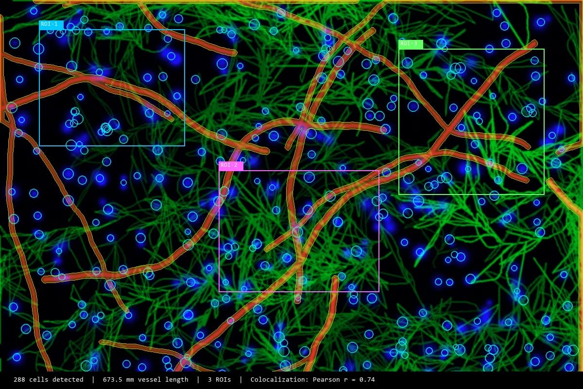
Cell Counting & Stereology
Unbiased cell population estimates using the Optical Fractionator with AI-assisted detection.

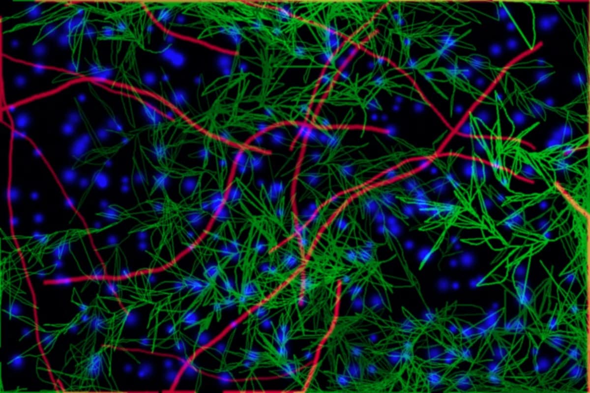
Vessel Reconstruction
3D vascular graph extraction with automated loop detection, diameter mapping, and large-volume support.

Fluorescence Colocalization
Quantify spatial overlap of fluorescent markers with Pearson’s, Manders’, and object-based methods.

Brain Atlas Registration
Register brain images to 30+ standardized atlases for automated region identification and cross-animal comparison.
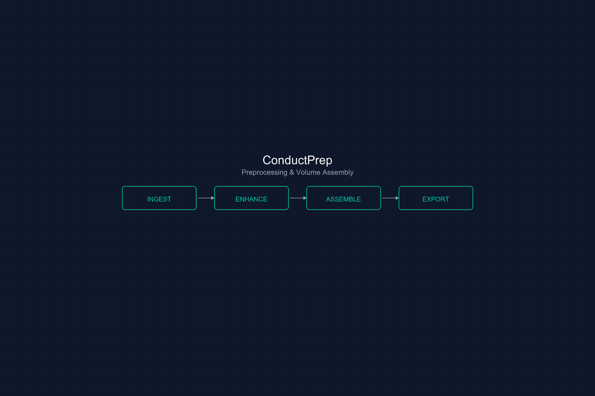
ConductPrep
Universal preprocessing — format conversion, tile stitching, deconvolution, and registration for any microscope.
Four steps from image to insight
How It Works
Import
Load Your Data
- TIFF, CZI, ND2 microscopy stacks
- Bio-Formats compatible images
- Drag-and-drop batch import
Define
Set Sampling
- Region of interest selection
- Stereological sampling grids
- Seed point placement
Detect
AI-Assisted Review
- AI suggests candidate cells
- Accept, reject, or refine
- Respects guard zones & counting rules
Export
Open Formats
- CSV count tables
- SWC morphology files
- VTK 3D meshes — no lock-in
Built for modern microscopy labs
No hardware dongle
Software-only licensing. No USB dongles, no node-locked seats. Move between workstations freely.
Works on your microscope
Microscope-agnostic. Import from any acquisition system — Zeiss, Leica, Nikon, Olympus — via Bio-Formats.
AI inside the probe
Detection runs inside the stereological sampling frame, respecting guard zones and counting rules. Not a generic counter.
Open export formats
CSV for counts, SWC for morphology, VTK for 3D meshes. Your data is yours.
Real-time statistics
Coefficient of Error updates live as you count. Stop when you’ve reached your confidence threshold.
Flexible licensing
Annual, multi-year, or perpetual options. Volume discounts for multi-seat labs.
From acquisition to publication
ConductVision Microscopy sits at the center of your imaging pipeline — from raw acquisition through quantification to publication-ready output.
Flexible plans for every lab
From single-workstation licenses to department-wide deployments. No hardware costs, no per-seat dongles.
Individual
Single workstation
- All microscopy modules
- Free software updates
- Standard support
Lab
Up to 5 seats
- All microscopy modules
- Free software updates
- Priority support
- Volume discount
Department
Unlimited seats + priority support
- All microscopy modules
- Free software updates
- Dedicated support
- Custom integration
Ready to modernize your microscopy pipeline?
Get reproducible quantification without the hardware lock-in.