
Equipping Your Histology Lab
From paraffin block to stained slide, the five instruments that turn tissue into data.
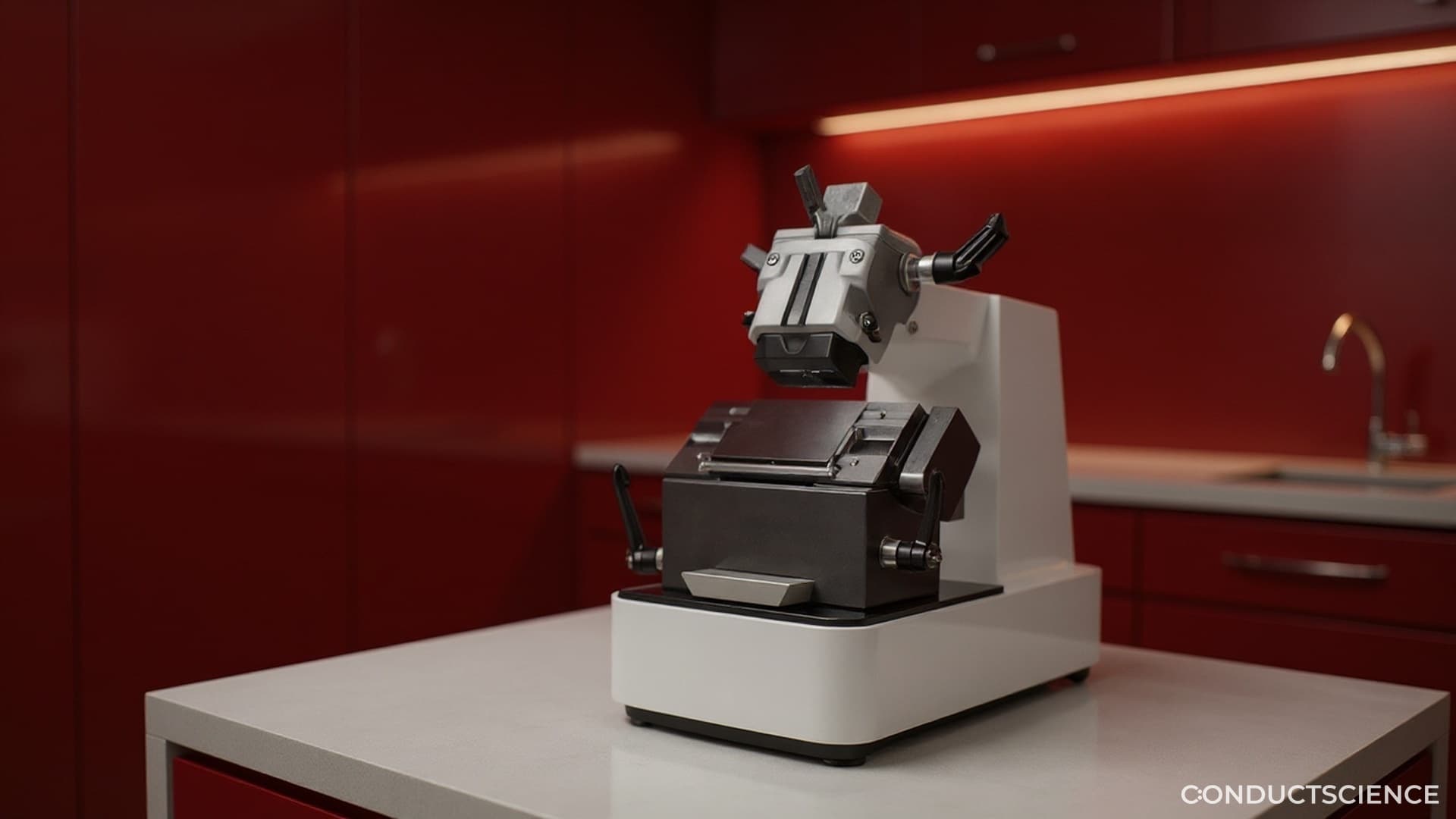
Rotary Microtome
Five microns. That is the thickness you need, thinner than a strand of spider silk, and the rotary microtome is the only thing in the lab that can deliver it on command. You clamp the paraffin block in the chuck, advance the wheel, and a ribbon of tissue uncoils onto the blade like a single long thought. The first section is too thick. The second tears at the edge. By the fifth, the ribbon glides off in perfect sheets. You float them on warm water and lift them onto slides. Three hours of trimming, an entire week of fixation, all of it built so this moment can happen cleanly.
Explore the Rotary Microtome→
Cryostat
Negative twenty Celsius inside the chamber, a small arctic on the bench. You snap-freeze the brain in OCT, mount it on the chuck, and bring the blade down through frozen tissue at ten microns per pass. There is no paraffin here, no antigen masking, no waiting overnight for processing. The slice you cut at 9:14 a.m. is on a slide and incubating with primary antibody by 9:30. Cryosectioning is for the experiments that cannot wait for chemistry to forget what the tissue looked like alive.
Explore the Cryostat→
Tissue Embedding Center
A liver biopsy enters the carousel at midnight in ten percent formalin. By morning it has passed through ascending ethanols, two changes of xylene, and three baths of molten paraffin, every step timed to the minute. You open the basket, lift the cassette into the embedding mold, orient the cut surface down, and pour wax around it. Thirty seconds on the cold plate and the block is solid. The tissue is now suspended in paraffin like a fly in amber, ready to wait years for the question that will need it.
Explore the Tissue Embedding Center→
Slide Dryer & Hot Plate
A freshly cut paraffin ribbon arrives on the slide curled and creased, a pale comma on glass. You lower it onto the hot plate at 60 Celsius and watch the wax soften, the section unfurl, the wrinkles smooth themselves out over thirty seconds. Then it dries flat overnight, the paraffin bonding the tissue to the slide so firmly that nothing in the staining bath can lift it off. Every hematoxylin streak you ever read at the microscope, every clean H&E in a published figure, started with a section that was dried this carefully and held this still.
Explore the Slide Dryer & Hot Plate→
Biological Microscope
Four objectives on the turret. The 4x for orientation, the 10x for architecture, the 20x for cell populations, the 40x for nuclei. You start at low power and move down the slide in a serpentine, the same way pathologists have done it for a century. Then a region catches your eye. You snap to 40x. The mitotic figure is unmistakable, suspended in metaphase. You photograph the field, drop a scale bar, and now the question that started as a hypothesis has its first piece of evidence.
Explore the Biological Microscope→Five instruments. One slide. Every diagnosis ever made started here.
Start Building Yours