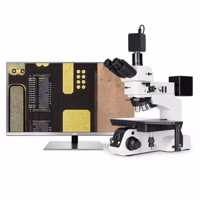
Polarizing Biological Microscope
Compound optical microscope with polarizing capability for analysis of birefringent materials and optically anisotropic structures in biological specimens.
| Automation Level | manual |
The Polarizing Biological Microscope is a compound optical instrument designed for quantitative analysis of birefringent materials in biological specimens. This microscope incorporates crossed polarizing filters that enable visualization and measurement of optically anisotropic structures such as collagen fibers, muscle striations, crystalline deposits, and organized cellular components. The polarization capability allows researchers to distinguish between isotropic and anisotropic materials based on their optical properties when placed between crossed polarizers.
The instrument features standard brightfield illumination with the addition of polarizing elements that can be engaged for specialized applications requiring birefringence analysis. The compact design with dimensions of 42.5 × 32.7 × 27.3 cm and 7.5 kg weight makes it suitable for routine laboratory use while maintaining the optical precision required for quantitative polarization microscopy in research and diagnostic applications.
How It Works
Polarization microscopy operates on the principle of birefringence, where certain materials split incident light into two orthogonal polarized rays that travel at different velocities through the specimen. The microscope incorporates a polarizer below the specimen stage and an analyzer above the objective lens, positioned at 90 degrees to each other (crossed polarizers). When polarized light encounters an optically isotropic material, it maintains its polarization state and is blocked by the crossed analyzer, appearing dark in the field of view.
Anisotropic materials, however, alter the polarization state of transmitted light due to their ordered molecular structure. This results in some light passing through the analyzer, causing birefringent structures to appear bright against a dark background. The degree of brightness depends on the material's birefringence strength and the orientation relative to the polarization axes. Rotating the specimen stage allows determination of extinction positions and measurement of optical properties such as retardation values.
The instrument's optical system maintains standard compound microscope functionality while adding the polarization components necessary for specialized analysis. This dual capability allows switching between conventional brightfield observation and polarization analysis within the same viewing session.
Features & Benefits
Automation Level
- manual
Research Domain
- Analytical Chemistry
- Cell Biology
- Clinical Diagnostics
- Developmental Biology
- Histopathology
- Materials Science
Weight
- 7.5 kg
Dimensions
- L: 42.5 mm
- W: 32.7 mm
- H: 27.3 mm
Comparison Guide
| Feature | This Product | Typical Alternative | Advantage |
|---|---|---|---|
| Optical Design | Compound microscope with integrated polarizing elements | Basic models often use simple polarizing filters without precision alignment | Ensures consistent optical performance and accurate extinction measurements across the field of view |
| Illumination System | Adjustable brightfield illumination with polarizer integration | Entry-level systems may lack proper Köhler illumination setup | Provides uniform field illumination essential for quantitative polarization analysis |
| Mechanical Stability | 7.5 kg stable construction with precision stage | Lighter instruments may lack mechanical stability for extended use | Maintains optical alignment during long observation sessions and precise angular measurements |
| Filter Quality | Precision-aligned polarizer and analyzer elements | Lower-cost models may use less precise filter mounting | Ensures accurate extinction and consistent birefringence measurements across specimens |
This polarizing biological microscope combines standard compound microscopy with precision polarization analysis capabilities in a stable, laboratory-suitable package. The instrument provides both qualitative and semi-quantitative analysis options for birefringent materials while maintaining compatibility with standard microscopy accessories and protocols.
Practical Tips
Verify extinction quality daily by checking field darkness with crossed polarizers and no specimen present.
Why: Proper extinction ensures accurate detection of weak birefringence and reliable quantitative measurements.
Clean polarizing filters monthly with lens cleaning solution and store with protective covers between uses.
Why: Contaminated or scratched polarizers introduce artifacts that compromise measurement accuracy and image quality.
Use mounting media with refractive index matched to your specimens to minimize edge artifacts.
Why: Index matching reduces optical distortion at specimen boundaries that can mask weak birefringence signals.
Record specimen orientation relative to stage markings for reproducible angular measurements.
Why: Consistent orientation reference enables comparison of birefringence data between samples and experimental sessions.
If extinction appears uneven across the field, check for mechanical stress in optical components and filter alignment.
Why: Uneven extinction indicates optical misalignment that will produce inconsistent birefringence measurements across the viewing area.
Adjust illumination intensity to minimum levels sufficient for observation to prevent specimen heating and degradation.
Why: Excessive illumination can alter specimen structure and introduce thermal stress artifacts in temperature-sensitive materials.
Allow the instrument to reach thermal equilibrium for 15 minutes before critical measurements.
Why: Temperature-induced mechanical stress can affect optical alignment and polarizer performance during precision analysis.
Setup Guide
What’s in the Box
- Polarizing biological microscope (typical)
- Polarizer filter assembly (typical)
- Analyzer filter assembly (typical)
- Standard eyepieces (typical)
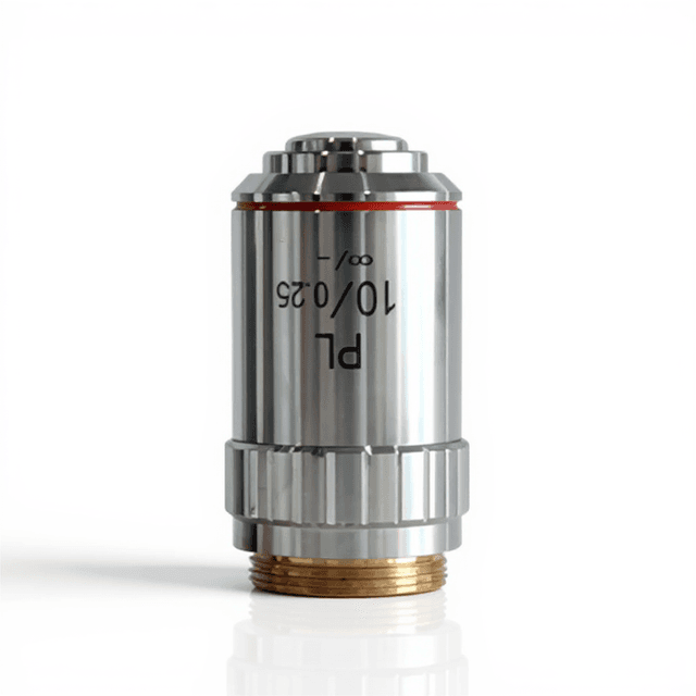
- Objective lens set (typical)
- Power cord (typical)
- Dust cover (typical)
- User manual (typical)
- Optical cleaning kit (typical)
Warranty
ConductScience provides a standard one-year manufacturer warranty covering defects in materials and workmanship. Technical support is available for optical alignment, maintenance procedures, and application guidance throughout the warranty period.
Compliance
What types of specimens are best suited for polarization microscopy analysis?
Specimens containing organized structures such as collagen fibers, muscle striations, crystalline deposits, cellulose, starch granules, and polymer materials exhibit strong birefringence. Fixed tissue sections should be cut at 5-10 μm thickness for optimal light transmission and contrast.
Can this microscope perform quantitative retardation measurements?
The basic polarizing setup allows qualitative assessment of birefringence and extinction angle determination. For quantitative retardation measurements, additional compensators or quarter-wave plates would need to be added to the optical path.
How do I achieve proper extinction for accurate measurements?
Cross the polarizer and analyzer until the field appears maximally dark with no specimen present. Fine-tune the analyzer position while observing the field brightness, and mark this position as your reference point for all subsequent measurements.
What is the recommended procedure for mounting specimens?
Use mounting media with refractive index close to 1.518 to minimize artifacts. Avoid thick coverslips that may introduce strain birefringence, and ensure specimens are completely flat to prevent optical distortion.
How does this compare to fluorescence microscopy for fiber analysis?
Polarization microscopy reveals intrinsic optical properties without requiring fluorescent labeling, preserving natural specimen structure. However, it is limited to birefringent materials, while fluorescence can target specific molecular components regardless of optical properties.
What maintenance is required for the polarizing elements?
Clean polarizing filters monthly with appropriate optical cleaning solutions and lint-free cloths. Check for scratches or stress-induced birefringence in the filters, which would compromise measurement accuracy. Store with protective covers when not in use.
Can I use oil immersion objectives with this polarizing setup?
Yes, oil immersion objectives are compatible, but ensure the immersion oil has minimal intrinsic birefringence. Use specialized low-stress immersion oils designed for polarization work to avoid introducing optical artifacts at high magnifications.
What factors affect image contrast in polarized light?
Contrast depends on specimen birefringence strength, thickness, orientation relative to polarizer axes, and illumination intensity. Optimal results require balancing these factors while maintaining extinction quality in the optical system.
Have a question about this product?
Accessories
Enhance your setup with compatible accessories

Replacement Parts & Consumables


Frequently Bought Together
Upgrade Options
Related Products