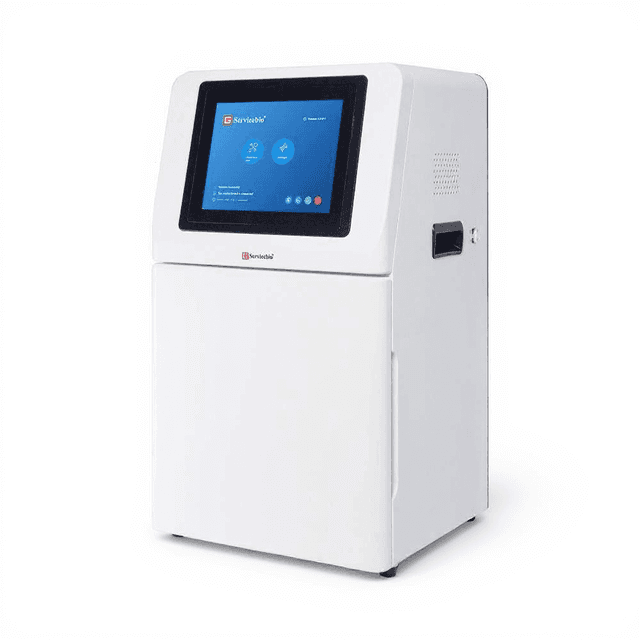
Automatic Gel Imaging System
Fully automated CCD-based gel imaging system with 5.03MP resolution, motorized optics, and integrated analysis software for nucleic acid and protein gel documentation.
| Instrument Type | Imaging Systems |
| Application Area | Protein Analysis |
| Automation Level | fully-automated |
| objective-lens-focal-length | 8-48mm |
| operating_wavelengths | 254nm, 302nm, 365nm, 590nm |
| exposure_time_range | 1ms-1000ms |
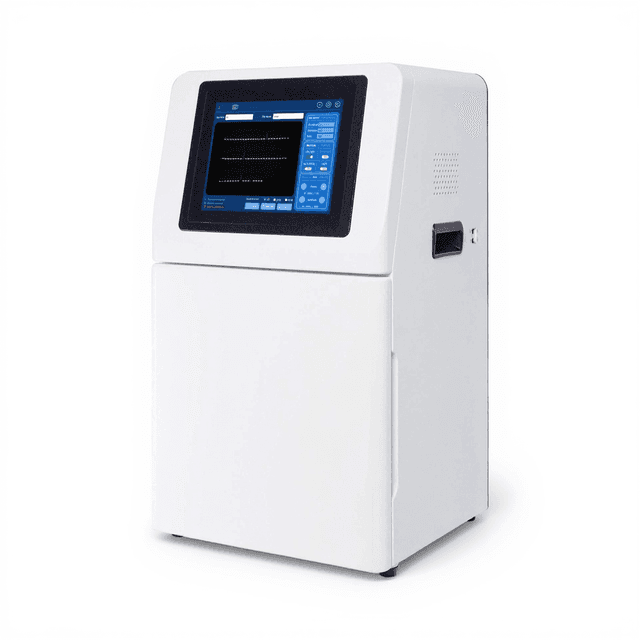
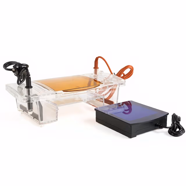
The Automatic Gel Imaging System (BIO-0020) provides high-resolution documentation and quantitative analysis of nucleic acid and protein separations following gel electrophoresis. This fully automated system integrates a 5.03 megapixel CCD camera with motorized lens control (8-48mm, F1.2), UV transillumination at 302nm, and white light illumination for comprehensive gel visualization. The system captures 16-bit images with >4.8 optical density range and quantum efficiency >65%, enabling detection of both high and low abundance targets.
The imaging chamber accommodates gels up to 21×21cm with automated focus adjustment and exposure control from 1-3000ms. Included software provides band detection, molecular weight estimation, and quantitative densitometry analysis. The system operates with multiple fluorescent dyes including ethidium bromide, SYBR Safe, and protein stains like Coomassie blue, supporting both UV and visible light applications in molecular biology workflows.
How It Works
The system employs CCD-based digital imaging to capture fluorescence or absorbance signals from stained nucleic acids and proteins separated by gel electrophoresis. UV transillumination at 302nm excites fluorescent intercalating dyes like ethidium bromide, producing emission wavelengths around 590nm that the high quantum efficiency CCD sensor detects with >65% efficiency. The 16-bit depth provides 65,535 gray levels for precise quantification across a >4.8 optical density range.
Motorized lens control (8-48mm focal length, F1.2 aperture) automatically adjusts magnification and focus based on gel size and band density. The darkroom hood eliminates ambient light interference while LED white light illumination enables visualization of visible stains like methylene blue or Coomassie brilliant blue. Software algorithms perform background subtraction, band detection, and molecular weight calculations by comparing migration distances to known DNA or protein standards.
Exposure times from 1-3000ms accommodate both bright fluorescent signals and weak chemiluminescent reactions. The 2592×1944 pixel resolution captures fine detail across the 21×21cm imaging area, enabling detection of bands separated by as little as 2-3mm on high-density gels.
Features & Benefits
Instrument Type
- Imaging Systems
Application Area
- Protein Analysis
Automation Level
- fully-automated
objective-lens-focal-length
- 8-48mm
operating_wavelengths
- 254nm, 302nm, 365nm, 590nm
exposure_time_range
- 1ms-1000ms
uv_wavelength_range
- 254nm-302nm
fluorescence_emission_wavelength
- 590nm
camera_type
- high-resolution CCD camera
autofocus_length
- 8-48mm
included_components
- darkroom hood, UV illuminator, CCD camera, computer software, white sample plate, UV sample plate, special filter for nucleic acid dye, overlay glue cutting filter
Pixel
- 2592×1944 (5.03MP)
Exposure Time
- 1ms~3000ms
QE Value
- >65%
Bit Depth
- 16 bit
OD
- ≥4.8OD
Lens
- Motorized 8~48mm, F1.2
Trans-UV
- 302nm
UV Area
- 21×21cm
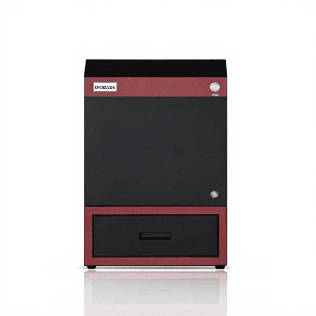
Dimensions
- 340mm x 475mm x 725mm
Research Domain
- Analytical Chemistry
- Cancer Research
- Cell Biology
- Environmental Monitoring
- Food Science
- Microbiology
- Molecular Biology
- Pharmaceutical QC
Power/Voltage
- 100~240V, 50/60Hz
Weight
- 31kg
Weight
- 68.34 lbs
Dimensions
- L: 8.27 in
- W: 8.27 in
- H: 44.8 in
Comparison Guide
| Feature | This Product | Typical Alternative | Advantage |
|---|---|---|---|
| Camera Resolution | 5.03 megapixels (2592×1944) | Entry-level systems often provide 1-3 megapixel resolution | Higher resolution enables detection of closely spaced bands and precise molecular weight determination on complex gels. |
| Dynamic Range | >4.8 optical density with 16-bit depth | Basic models typically offer 12-bit depth with limited OD range | Captures both faint and intense bands simultaneously without saturation, reducing need for multiple exposures. |
| Lens System | Motorized 8-48mm zoom with F1.2 aperture | Fixed focal length lenses or manual zoom systems | Automated magnification control optimizes image quality for different gel sizes without manual adjustment. |
| Illumination Options | Integrated UV (302nm) and white LED systems | Single illumination type or separate light sources | Supports both fluorescent and visible stains without external equipment changes or filter mounting. |
| Imaging Area | 21×21cm field of view | Smaller systems often limited to 15×15cm or single mini-gel format | Accommodates large format gels and multiple samples for higher throughput analysis. |
| Exposure Control | 1-3000ms variable exposure time | Limited exposure range or manual control only | Automated exposure optimization for both rapid fluorescence and extended chemiluminescence applications. |
The system provides research-grade CCD imaging with 5.03 megapixel resolution, 16-bit dynamic range, and integrated dual illumination. The motorized optics and large imaging area support diverse gel formats while maintaining quantitative accuracy for molecular biology applications.
Practical Tips
Run molecular weight standards on each gel and calibrate the software sizing algorithm before analyzing unknown samples.
Why: Migration rates vary with gel concentration, buffer conditions, and voltage, requiring fresh calibration for accurate molecular weight determination.
Use the lowest exposure time that provides adequate signal to minimize noise and prevent CCD saturation in bright regions.
Why: Shorter exposures reduce thermal noise while the high quantum efficiency sensor maintains sensitivity for weak signals.
Keep the imaging chamber and sample plates clean using appropriate solvents to prevent fluorescent contamination between samples.
Why: Residual dye or protein deposits create background fluorescence that reduces image quality and quantification accuracy.
Save images in 16-bit format for quantitative analysis even if 8-bit display appears adequate for visual inspection.
Why: Quantitative densitometry requires the full dynamic range to accurately measure band intensities across multiple orders of magnitude.
Always use the darkroom hood when imaging UV-excited samples to prevent eye exposure to 302nm radiation.
Why: UV wavelengths below 320nm can cause corneal and retinal damage even at low intensities used for gel transillumination.
If bands appear oversaturated, reduce exposure time rather than adjusting illumination intensity to maintain uniform lighting.
Why: Changing illumination affects the entire imaging field while exposure control selectively prevents saturation in bright regions.
Image gels immediately after staining to prevent dye migration or photobleaching that reduces band sharpness.
Why: Many fluorescent dyes are light-sensitive and DNA bands continue migrating slowly even after electrophoresis completion.
Use multiple exposure times for highly variable samples to ensure optimal signal capture across all bands of interest.
Why: Single exposure may oversaturate strong bands or underexpose weak signals, while multiple exposures enable software-based composite imaging.
Setup Guide
What’s in the Box
- Automatic gel imaging system with CCD camera
- Darkroom hood assembly
- UV illuminator (302nm)
- CCD camera module
- Analysis software package
- White sample plate
- UV sample plate
- Special filter for nucleic acid dye
- Overlay glue cutting filter
- Power supply cable
- USB interface cable
- User manual and software guide
Warranty
ConductScience provides a standard one-year manufacturer warranty covering parts and labor, with technical support for software configuration and operational guidance.
Compliance
References
Background reading relevant to this product:
What is the minimum detectable DNA concentration on agarose gels?
Detection limits depend on gel thickness, dye type, and band size. With ethidium bromide staining, typical detection ranges from 5-10ng per band. The >4.8 OD range and 16-bit depth enable detection across 4+ orders of magnitude concentration range.
Can the system image protein gels stained with Coomassie blue?
Yes, the integrated white LED illumination and included white sample plate support visible stains including Coomassie brilliant blue, silver stains, and methylene blue. The broad spectral response of the CCD camera captures visible wavelength absorption.
What gel sizes are compatible with the imaging area?
The 21×21cm imaging area accommodates standard large format gels (20×20cm), multiple mini-gels (8×10cm), or single medium gels (15×15cm). The motorized lens adjusts magnification automatically for optimal resolution.
Does the software perform molecular weight calculations?
Yes, the analysis software includes molecular weight estimation based on migration distance comparison to standard DNA or protein ladders. Users input ladder specifications and the software generates standard curves for unknown band sizing.
How does the system handle chemiluminescent Western blots?
The high quantum efficiency CCD sensor (>65%) and extended exposure capability (up to 3000ms) enable chemiluminescent detection. The darkroom hood eliminates ambient light interference during long exposures.
What file formats are supported for image export?
Consult product datasheet for specific file format specifications. Most gel imaging systems support TIFF, JPEG, and proprietary formats for quantitative analysis while maintaining 16-bit depth for research applications.
Can multiple users access the system simultaneously?
The system connects via USB to a single computer workstation. Multiple user access would require network configuration of the host computer rather than direct instrument sharing.
What maintenance is required for the CCD camera?
CCD sensors require minimal maintenance but should be kept dust-free and operated within specified temperature ranges. The darkroom hood protects the camera from laboratory contaminants during routine use.
Have a question about this product?
Accessories
Enhance your setup with compatible accessories