Integrated Automatic Gel Imaging System
Dark field gel imaging system with 6.0 MP camera, 302nm UV/LED illumination, and integrated touchscreen for comprehensive nucleic acid documentation and quantitative analysis.

Louise Corscadden, PhD
Director of Science · ConductScience
Ask Louise about Integrated Automatic Gel Imaging System fit, setup, configuration, or quote prep.
Already working with us? Sign in to connect this with My Scientist.
Key Specifications
Full details →- Model fit
- Configured during quote
- SKU family
- BIO-BK-AGX3
- Sizing
- 8.27 x 8.27 x 38.0 cm
- Ordering
- Online checkout and quote request available
- Category
- Gel & Clarity Testing
- Build notes
- Confirm accessories, station layout, and support needs before purchase
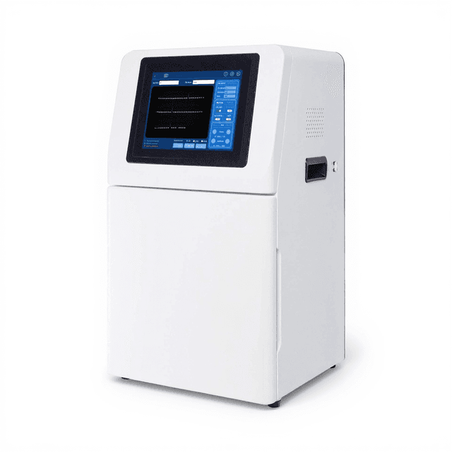
The Integrated Automatic Gel Imaging System (BIO-BK-AGX3) is a dark field imaging platform designed for comprehensive nucleic acid documentation across multiple fluorescent dye systems. The system combines a 6.0 MP camera (2592×1944 pixels) with 16-bit pixel depth and >79% photosensitive quantum efficiency for quantitative gel analysis. A motorized autofocus lens (8-48mm, F1.2) provides optical resolution across a 21×21 cm imaging area with automated focusing capabilities.
The integrated 12.5-inch touch HD display enables direct system control without external computer requirements. Standard 302 nm UV transillumination supports ethidium bromide and SYBR-based fluorescent dyes, while LED white light illumination accommodates visible stains including methylene blue and Coomassie blue. The multilayer coated filter system (590nm standard) provides spectral selectivity with ≥4.8 OD optical density for background suppression. Exposure times from 1ms to 3000ms accommodate varying sample concentrations and dye intensities, while 20 million pixel accuracy ensures precise band detection and quantification.
How It Works
The system operates through dark field imaging principles where nucleic acids are visualized using fluorescent dye intercalation or binding. When excited by 302nm UV light, fluorescent dyes such as ethidium bromide, SYBR Green, or SYBR Safe emit visible light at characteristic wavelengths. The 590nm multilayer coated filter selectively transmits fluorescent emission while blocking excitation wavelengths, creating high-contrast images with ≥4.8 OD background suppression.
The 6.0 MP camera with >79% quantum efficiency captures emitted fluorescence across the 21×21 cm imaging area. The motorized F1.2 lens system automatically adjusts focus from 8-48mm focal length to optimize resolution for different gel thicknesses and sample types. Exposure times ranging from 1ms to 3000ms allow optimization for varying dye concentrations and DNA amounts. For visible stains, LED white light illumination provides uniform transillumination without fluorescent excitation.
The integrated 12.5-inch touch HD display processes images in real-time using 16-bit pixel depth (0-65535 intensity range) for quantitative band analysis. The dark field configuration eliminates ambient light interference, ensuring consistent documentation across different laboratory lighting conditions. USB connectivity enables data transfer and external storage of high-resolution gel images with 20 million pixel accuracy for subsequent analysis.
Features & Benefits
camera_type
- dark field camera module
display_size
- 12.5 inches
filter_type
- multilayer coated filter
connectivity
- USB port
compatible_dyes
- EB, SYBR Gold, SYBR Green, SYBR Safe, Gel Red Gel, Green Texas Red, Fluorescein labeled DNA/RNA
Automation Level
- semi-automated
Pixel
- 2592×1944 (5.03MP)
Exposure Time
- 1ms~3000ms
QE Value
- >65%
Bit Depth
- 16 bit
OD
- ≥4.8OD
Lens
- Motorized 8~48mm, F1.2
Trans-UV
- 302nm
UV Area
- 21×21cm
Accuracy
- 20 million
Display Type
- Touch HD
Research Domain
- Analytical Chemistry
- Cancer Research
- Cell Biology
- Clinical Diagnostics
- Environmental Monitoring
- Food Science
- Microbiology
- Pharmaceutical QC
Power/Voltage
- 100~240V, 50/60Hz
Weight
- 31kg
Weight
- 68.34 kg
Dimensions
- L: 8.27 mm
- W: 8.27 mm
- H: 38.0 mm
| Feature | This Product | Typical Alternative | Advantage |
|---|---|---|---|
| Camera Resolution | 6.0 MP (2592×1944 pixels) | Entry-level systems often provide 3-5 MP cameras | Higher resolution enables detection of smaller bands and more precise quantitative analysis |
| Quantum Efficiency | >79% photosensitive efficiency | Basic systems typically offer 60-70% efficiency | Superior light sensitivity reduces exposure times and detects low-abundance samples |
| Illumination Options | Dual system: 302nm UV + LED white light | Many systems offer single illumination mode | Flexibility to use fluorescent or visible stains without equipment changes |
| Display Interface | Integrated 12.5-inch touch HD display | Most systems require external computer connection | Standalone operation reduces bench space requirements and setup complexity |
| Lens System | Motorized autofocus 8-48mm, F1.2 | Manual focus systems with fixed focal length | Automatic optimization eliminates focus adjustment time and user variability |
| Filter Specifications | 590nm multilayer coated with ≥4.8 OD | Standard filters often provide 3-4 OD background suppression | Enhanced background suppression improves signal-to-noise ratio for low-intensity bands |
The system combines high-resolution imaging with dual illumination capabilities and standalone operation through the integrated touchscreen interface. The motorized autofocus lens and high quantum efficiency camera provide technical advantages for quantitative gel analysis applications.
Practical Tips
Perform weekly focus calibration using a standard test gel or calibration slide to maintain optimal image sharpness across the 21×21 cm area.
Why: Regular calibration ensures consistent focus quality and compensates for any mechanical drift in the motorized lens system.
Clean the UV transilluminator surface weekly with 70% ethanol and lint-free wipes to remove gel residue and maintain uniform illumination.
Why: Gel residue and fingerprints can create uneven illumination patterns that affect image quality and quantitative analysis.
Use the shortest exposure time that provides adequate signal to avoid saturation of bright bands while maintaining detection of weak bands.
Why: Optimal exposure maximizes the dynamic range for quantitative analysis and prevents loss of linear response in high-intensity regions.
Include molecular weight markers and loading controls in each gel for band size determination and loading normalization.
Why: Reference standards enable accurate molecular weight estimation and account for loading variations between samples.
Always use UV-protective eyewear when working with the 302nm transilluminator and ensure proper room ventilation when using ethidium bromide.
Why: UV exposure can cause eye damage, and ethidium bromide vapors pose health risks requiring appropriate safety precautions.
If autofocus fails to converge, manually clean the camera lens and ensure the gel surface is free of bubbles or debris.
Why: Surface contamination or optical obstructions can interfere with the autofocus system's ability to detect the focal plane accurately.
Save images in TIFF format for quantitative analysis to preserve full dynamic range and avoid compression artifacts.
Why: TIFF format maintains the complete 16-bit pixel depth necessary for accurate densitometry and comparative analysis.
Monitor UV tube output periodically using a UV meter to ensure consistent 302nm intensity for reproducible fluorescent excitation.
Why: UV tube degradation over time can reduce excitation efficiency and affect the sensitivity and consistency of fluorescent detection.
Setup Guide
What’s in the Box
- Integrated gel imaging system main unit
- Power cord (100-240V)
- 590nm fluorescent filter (installed)
- USB cable
- User manual and quick start guide
- Calibration reference materials (typical)
- Software installation media (typical)
- Protective camera lens cover (typical)
Warranty
ConductScience provides a 1-year manufacturer warranty covering parts and labor, with technical support for installation and operational guidance. Extended service plans available for ongoing maintenance and calibration services.
Compliance
References
Background reading relevant to this product:
What fluorescent dyes are compatible with the 590nm filter system?
The 590nm filter accommodates EB, SYBR Gold, SYBR Green, SYBR Safe, Gel Red, Texas Red, and fluorescein-labeled DNA/RNA. For optimal results with other dyes, consult the spectral emission characteristics against the filter specifications.
Can the system quantify DNA band intensities for comparative analysis?
Yes, the 16-bit pixel depth (0-65535 range) with 20 million pixel accuracy enables precise densitometry measurements. Variable exposure times (1-3000ms) allow optimization for linear quantification ranges.
What gel sizes and formats are supported by the imaging area?
The 21x21 cm imaging area accommodates standard mini-gels, midi-gels, and large format gels up to 20x20 cm. Multiple smaller gels can be imaged simultaneously within the field of view.
How does the autofocus system work with different gel thicknesses?
The motorized F1.2 lens automatically adjusts focus from 8-48mm focal length based on gel surface detection. The system compensates for gel thickness variations from 0.5mm to 15mm typical range.
What maintenance is required for the UV transilluminator?
Regular cleaning of the UV surface with ethanol and monitoring of 302nm output intensity. UV tube replacement typically required after 2000-5000 hours of operation depending on usage patterns.
Can visible stains be documented without the fluorescent filter?
Yes, the LED white light system accommodates visible stains like methylene blue and Coomassie blue. Remove the 590nm filter for direct visible light imaging without fluorescent excitation.
What image file formats are supported for data export?
The system supports standard formats including TIFF, JPEG, and BMP through USB connectivity. Consult the product datasheet for complete file format specifications and metadata options.
How does the system compare to traditional film-based documentation?
Digital documentation provides immediate results, quantitative analysis capabilities, and elimination of darkroom processing. The 6.0 MP resolution with >79% quantum efficiency often exceeds film sensitivity for fluorescent applications.
Can this system be used for western blot imaging, and does it produce digital figures?
Yes. For western blots stained with Coomassie Blue, Ponceau S, or other visible dyes, use the LED white light mode with the 590nm filter removed. The system captures high-resolution images and exports them as TIFF, JPEG, or BMP files, suitable for publication-ready figures and quantitative band densitometry. For chemiluminescent (ECL) detection, please confirm compatibility with your specific reagents and imaging conditions before use.
Have a question about this product?
Have a question? Just ask.
Send it over and we'll email you a personalized answer — no call, no scheduling.
Prefer to talk it through?
Accessories
Enhance your setup with compatible accessories