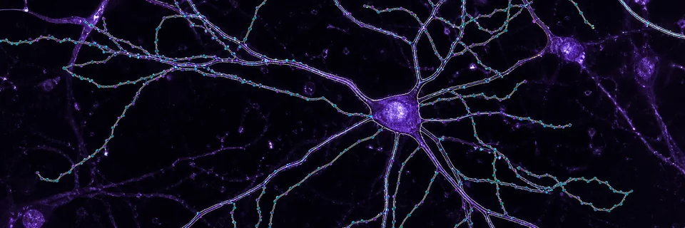
Neuroscience image analysis, from single neuron to whole-brain atlas.
Start with the measurement you need — traced neurons, spine turnover, glial activation, stereology counts, vasculature, colocalization, or atlas-mapped regions — and open the ConductVision workflow that produces it. These tools already exist; this page connects them.
- 9
- Neuroscience workflows, from single-neuron morphology to whole-brain atlas registration
- 2
- Method families: neuron and glial morphology, plus quantitative microscopy
- One engine
- Shared ConductVision segmentation, tracing, and batch export across every tool
Find the neuroscience workflow first
Image analysis is the measurement engine. Neuroscience is the application layer: neuron and glial structure, dendritic spines, unbiased counts, vasculature, and region mapping. This page groups those overlapping workflows in one place while keeping the existing ConductVision Morphology and Microscopy pages exactly where they are.
Neuron and glial structure
Reconstruct, measure, and compare cell morphology — from a single traced neuron to cohort-scale glial activation and dendritic-spine turnover.
Trace — neuron reconstruction
Reconstruct neurons from raw microscopy volumes to publication-ready SWC, with 40+ built-in morphology metrics on a single desktop.
Spine Dynamics
ML-assisted 4D analysis that aligns, tracks, and classifies dendritic spines across days or weeks — built for LTP/LTD, neurodegeneration, and drug-discovery studies.
Morphology Explorer
Explore neuronal morphology in interactive 3D — compare populations, visualize Sholl analysis, overlay segments, and export publication-ready figures.
Glial Morphometry
Quantify microglia ramification, astrocyte territory, and oligodendrocyte process complexity, with per-cell activation classes and cohort-scale batch comparison.
Quantitative microscopy
Turn microscopy stacks into rigorous measurements: unbiased counts, vascular graphs, colocalization statistics, atlas-mapped regions, and preprocessing at terabyte scale.
Stereology
Unbiased optical-fractionator counting with machine-learning detection and real-time CE tracking — statistically rigorous and hardware-independent.
Vessel — vascular reconstruction
Convert confocal and light-sheet stacks into flow-ready vascular graphs with automated loop detection, diameter mapping, and large-volume reconstruction.
Colocalization
Quantitative co-occurrence for multi-channel fluorescence — Pearson r, Manders M1/M2, Costes auto-threshold, and ROI-based batch statistics across cohorts.
Brain-Atlas registration
Map microscopy data to Allen CCFv3, Waxholm Rat, or custom atlases with affine plus nonlinear warp and per-region quantification.
Prep — microscopy preprocessing
Convert 150+ microscope formats, auto-stitch tiles, deconvolve, and process terabyte datasets before analysis.
Related ConductVision centers
Use Neuroscience as the entry point when you start with the brain. Use these centers when you already know the method or start from a different domain.
Life Science
Start here when the entry point is a cell, tissue, or assay: confluence, viability, histology, and fluorescence workflows.
Materials Science
Metallography, surfaces, particles, SEM, and CT — calibrated measurements and reviewable overlays for materials imaging.
Morphology center
The full morphology method hub: neuron tracing, spine dynamics, Sholl analysis, and ML classification.
Microscopy center
The full microscopy method hub: stereology, vessel reconstruction, colocalization, and atlas registration.
Need a neuroscience workflow mapped to your images?
Send representative stacks and the measurement you need. ConductScience can confirm the closest ConductVision workflow or scope a custom analysis path.