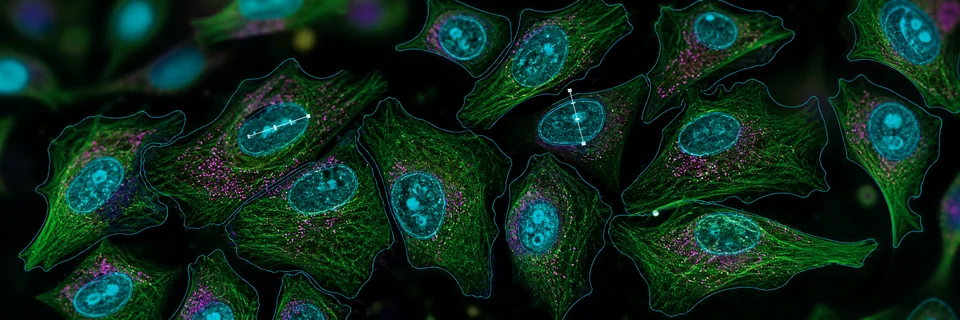
Life science image analysis for cells, tissue, assays, and microscopy.
Start with the biological sample or assay, then open the closest ConductVision workflow. Cell counting, confluence, viability, histology, fluorescence, microscopy, and morphology stay connected without replacing their existing pages.
- 17+
- life science entry points
- 5
- application families
- 0
- existing URLs replaced
Find the biology workflow first
Image analysis is the measurement engine. Life science is the application layer: cells, tissue, assays, microscopy, and morphology. This page groups overlapping biology workflows in one place while preserving the current ConductVision Image, Microscopy, Morphology, and Materials URLs.
Materials science remains its own atlas for metallography, surfaces, particles, textiles, electronics, geology, and inspection. Shared methods like segmentation, counting, thresholding, and batch export still use the same ConductVision engine.
Cell assays
Counts, confluence, viability, colonies, and plate-level readouts for routine cell biology.
Fluorescence markers
Nuclei, live/dead markers, multi-channel fields, and marker-positive populations.
Tissue and pathology
H&E nuclei, DAB staining, positive area, tissue regions, and histology exports.
Culture models
Scratch assays, spheroids, organoids, angiogenesis tubes, and time-lapse measurements.
Microscopy and morphology
Stereology, colocalization, vessel analysis, atlas registration, tracing, and morphometry.
Life science application atlas
Search the biology workflows by sample, stain, assay, modality, or output. Some cards open existing ConductVision Image pages, and others open the current microscopy, morphology, or bridge application pages.
 Fluorescence markers
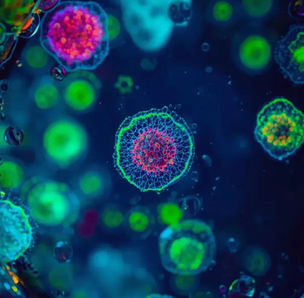
Automated Cell Counting (Fluorescence)
Precise cell counting using fluorescent markers
 Cell assays
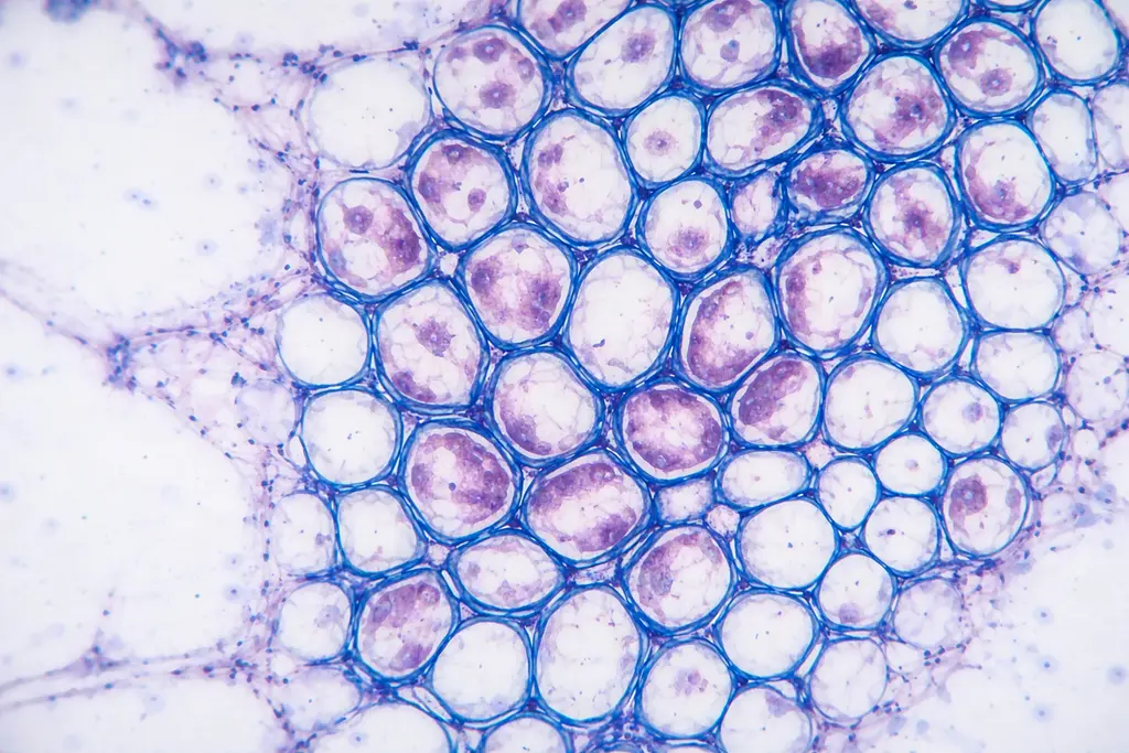
Cell Confluence
Measure culture confluence from phase-contrast images
 Fluorescence markers
Nuclei (Fluorescence)
Fluorescence-based nuclei detection and quantification
 Cell assays
Live/Dead Cell Viability
Cell viability counting from live/dead fluorescence images
 Cell assays
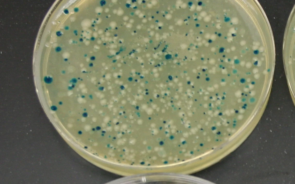
Colony Counting Software
AI colony counts from uploaded agar plate images
 Tissue and pathology
Hematoxylin-Stained Nuclei
Detect nuclei in H&E histology sections
 Tissue and pathology
DAB Stained Tissue Analysis
Quantify DAB staining intensity in IHC samples
 Tissue and pathology
Percent Area (General)
Coverage and area percentage calculations
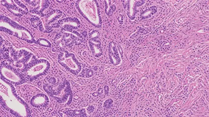 Tissue and pathology
Pathology & histology
Region and feature quantification on stained tissue sections.
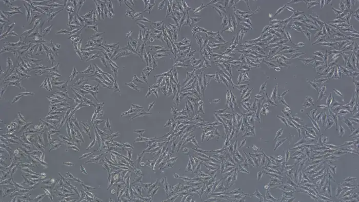 Cell assays
Cell counting & confluence
Counts, density, and confluence from brightfield and phase contrast.
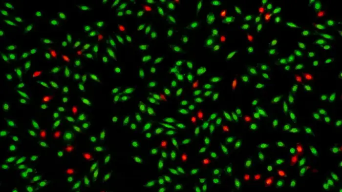 Cell assays
Viability & assays
Live and dead segmentation, viability ratios, and dose response.
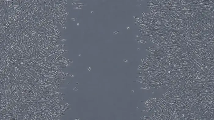 Culture models
Scratch & wound assay
Wound area and closure over time from migration assays.
 Culture models
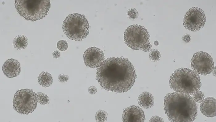
Organoid & spheroid
Size, count, and morphology of 3D culture structures.
 Culture models
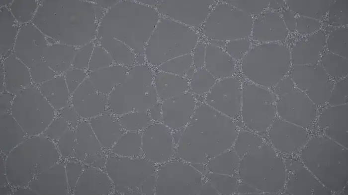
Angiogenesis & tubes
Network length, branch points, and node counts from tube-formation assays.
 Fluorescence markers
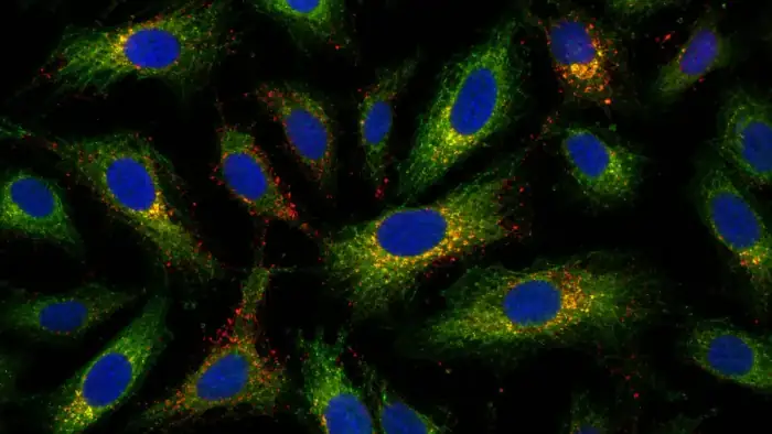
Fluorescence colocalization
Pearson, Manders, and Costes across multi-channel fields.
 Microscopy and morphology
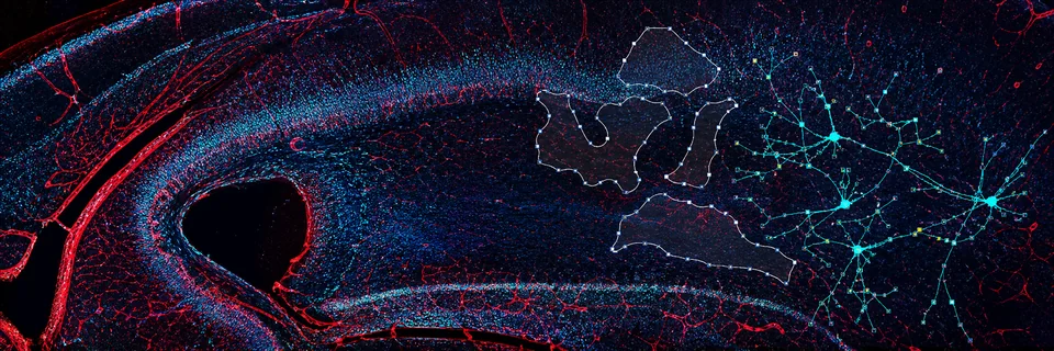
Microscopy analysis center
Stereology, atlas registration, vessel reconstruction, colocalization, prep, and spine workflows.
 Microscopy and morphology
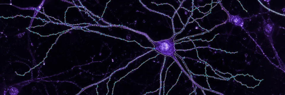
Morphology analysis center
Neuron tracing, morphology exploration, and glial morphometry for cell shape analysis.
Start with the assay category
These pages organize Life Science by how the lab describes the experiment. Each page links to existing ConductVision workflows and includes a meeting request path for custom assays.
Cell assay image analysis for counting, confluence, viability, and colonies.
Quantify routine cell biology images without changing the assay your lab already runs.
Tissue and pathology image analysis for stained sections and IHC.
Turn stained tissue images into counts, areas, intensity measurements, and reviewable maps.
Organoid and spheroid image analysis for 3D culture studies.
Measure 3D culture growth and morphology across populations, not just a few hand-picked structures.
Migration, wound healing, and angiogenesis image analysis.
Measure movement, closure, and network formation from assay images and time-lapse series.
Fluorescence microscopy image analysis for nuclei, markers, and colocalization.
Quantify markers, channels, nuclei, and colocalization while keeping overlays reviewable.
High-content image analysis for plate-based biological screening.
Scale image analysis from single fields to multi-well biological screens.
Additional application pages
These are net-new ConductVision Life Science pages for assays where the public search intent is strong and the current site does not already have a dedicated canonical page.
Application: Neurite outgrowth image analysis for neuronal culture assays
Measure neurite length, branching, and network growth from neuronal culture images.
Application: Cell painting image analysis for phenotypic profiling
Extract marker and morphology features from multiplex cell painting images.
Application: Transfection efficiency image analysis for reporter-positive cells
Calculate transfection efficiency from reporter and nuclear channels.
Application: ELISpot counting image analysis for spot-forming units
Count ELISpot wells consistently and export spot-level measurements.
Application: Plaque assay counting image analysis for viral plaques
Count plaques and quantify plaque area from plate or well images.
Application: Apoptosis assay image analysis for marker-positive cells
Quantify apoptosis markers and nuclear morphology from fluorescence images.
Application: Cell cycle image analysis for nuclear intensity and proliferation markers
Measure nuclear intensity and proliferation-marker readouts from microscopy images.
Application: Microsphere image analysis for drug delivery, encapsulation, and microcarrier beads
Size a whole microsphere batch from micrographs instead of hand-measuring a few dozen beads.
Related ConductVision centers
Use Life Science as the entry point when the user starts with biology. Use the centers below when the user already knows the technical method or adjacent domain.
ConductVision Image
The shared image-analysis engine behind cell, tissue, colony, particle, and material measurements.
Microscopy
Method-specific pages for stereology, colocalization, atlas registration, vessel analysis, and prep.
Morphology
Tracing and morphometry pages for neurons, glia, and cell-shape workflows.
Materials Science
The materials atlas stays separate for metallography, surfaces, powders, textiles, and inspection.
Need a life science workflow mapped to your assay?
Send representative images and the measurement you need. ConductScience can confirm the closest ConductVision workflow or scope a custom analysis path.