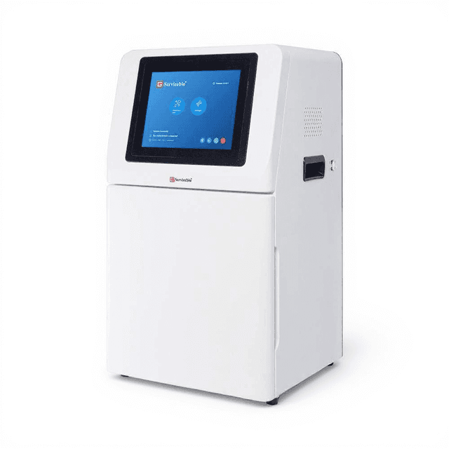
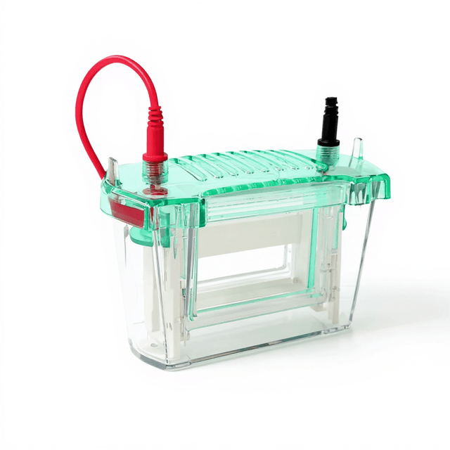
High sensitive Gel Imaging System
Fully automated gel documentation system with 6.3MP CCD camera and multi-wavelength illumination for high-resolution imaging of DNA, RNA, and protein electrophoresis gels.

Louise Corscadden, PhD
Director of Science · ConductScience
Ask Louise about High sensitive Gel Imaging System fit, setup, configuration, or quote prep.
Already working with us? Sign in to connect this with My Scientist.
Key Specifications
Full details →- Model fit
- Configured during quote
- SKU family
- BS-SCG-W1000
- Sizing
- 28.54 x 18.7 x 13.39 cm
- Ordering
- Online checkout and quote request available
- Category
- Gel & Protein Analysis
- Build notes
- Confirm accessories, station layout, and support needs before purchase
The BS-SCG-W1000 High Sensitive Gel Imaging System is a fully automated gel documentation platform designed for high-resolution visualization and analysis of nucleic acids and proteins separated by electrophoresis. The system integrates a high-resolution CCD camera (6.3 million pixels, 3072×2048 resolution) with multi-wavelength illumination capabilities, providing comprehensive imaging across UV (254nm, 302nm) and visible (365nm, 590nm) wavelengths for diverse staining methods including ethidium bromide, SYBR dyes, and protein stains.
Equipped with an 8-48mm autofocus objective lens and exposure time control from 1ms to 1000ms, the system accommodates samples ranging from single DNA bands to complex protein patterns on both agarose and polyacrylamide gels. The integrated darkroom hood eliminates ambient light interference, while specialized sample plates and filters optimize detection for both nucleic acid and protein applications, making it suitable for routine gel documentation, quantitative analysis, and research documentation requirements.
How It Works
The gel imaging system operates through controlled illumination and high-sensitivity charge-coupled device (CCD) detection. Sample fluorescence or absorption is excited by specific wavelengths (254nm and 302nm UV for nucleic acid dyes, 365nm and 590nm for protein stains and visible dyes), with emitted light captured by the 6.3 million pixel CCD sensor. The 2.4μm pixel size and low readout noise (2.14e-) enable detection of weak fluorescent signals and precise band quantification.
The autofocus objective lens system (8-48mm focal length range) automatically optimizes focus across the gel surface, while the integrated darkroom hood eliminates background illumination. Specialized sample plates provide optimal contrast for different gel types - white plates enhance protein band visibility in SDS-PAGE applications, while UV-transparent plates maximize nucleic acid fluorescence transmission. The exposure time control (1ms to 1000ms) accommodates samples with varying signal intensities, from bright ethidium bromide staining to weak protein bands.
Features & Benefits
Instrument Type
- Imaging Systems
Application Area
- Protein Analysis
Automation Level
- fully-automated
Cameras
- Black and White Camera
objective-lens-focal-length
- 8-48mm
operating_wavelengths
- 254nm, 302nm, 365nm, 590nm
exposure_time_range
- 1ms-1000ms
camera_type
- High-resolution CCD camera
camera_focus
- Autofocus length
objective_lens_focal_length
- 8-48mm
uv_wavelength_range
- 254nm-302nm
fluorescent_emission_wavelength
- 590nm
sample_plates
- White sample plate for SDS-PAGE, UV sample plate for nucleic acid gel
filters
- Special filter for nucleic acid dye, overlay glue cutting filter
Dimensions
- 475mm x 340mm x 725mm
Research Domain
- Analytical Chemistry
- Cancer Research
- Cell Biology
- Microbiology
- Molecular Biology
- Pharmaceutical QC
Weight
- 54.83 kg
Dimensions
- L: 28.54 mm
- W: 18.7 mm
- H: 13.39 mm
| Feature | This Product | Typical Alternative | Advantage |
|---|---|---|---|
| Camera Resolution | 6.3 million pixels (3072×2048) | Entry-level models typically offer 2-5 million pixels with lower resolution arrays | Higher pixel count enables detection of smaller bands and more accurate quantification of closely spaced samples |
| Wavelength Options | Four wavelengths: 254nm, 302nm, 365nm, 590nm | Basic systems often provide only 1-2 UV wavelengths | Multiple wavelengths accommodate diverse staining protocols without requiring separate imaging equipment |
| Focus System | Autofocus with 8-48mm focal length range | Manual focus systems require operator adjustment | Automated focusing reduces operator variability and ensures consistent image quality across different gel types |
| Exposure Control | Variable 1ms to 1000ms exposure time | Fixed or limited exposure options in basic models | Wide exposure range optimizes signal capture for both bright and weak fluorescent samples |
| Sample Plate Options | Specialized white and UV-transparent plates included | Single plate type or plates sold separately | Application-specific plates maximize contrast and signal transmission for different gel types |
| System Integration | Fully automated with computer integration | Semi-automated systems requiring more manual operation | Streamlined workflow reduces hands-on time and improves documentation consistency |
The BS-SCG-W1000 combines high-resolution imaging, multi-wavelength capability, and automated operation to provide comprehensive gel documentation functionality. The 6.3 million pixel CCD camera and four-wavelength illumination system accommodate diverse research applications from basic DNA analysis to complex protein studies.
Practical Tips
Verify focus accuracy monthly using a resolution test target placed at different positions on the sample plate.
Why: Autofocus systems can drift over time, affecting image sharpness and quantification accuracy.
Clean the sample plates weekly with appropriate solvents and inspect for scratches that could affect light transmission.
Why: Contaminated or damaged plates create artifacts and reduce image quality, particularly for UV applications.
Use the 60-80% dynamic range for quantitative work by adjusting exposure time to avoid pixel saturation.
Why: Saturated pixels cannot provide accurate intensity measurements, compromising quantitative analysis results.
Match sample plate selection to application - white plates for protein gels, UV-transparent for nucleic acids.
Why: Proper plate selection maximizes signal contrast and transmission efficiency for different detection methods.
If bands appear out of focus across the gel, recalibrate the autofocus system and check for gel warping.
Why: Uneven focus typically indicates mechanical misalignment or sample preparation issues rather than camera problems.
Include molecular weight standards and loading controls in every gel for normalization and quality assessment.
Why: Standards enable accurate size determination and loading control verification for quantitative comparisons.
Always close the darkroom hood before activating UV illumination to prevent eye and skin exposure.
Why: UV wavelengths (254nm, 302nm) can cause tissue damage even with brief exposure times.
Monitor UV lamp intensity monthly and replace according to manufacturer specifications to maintain consistent excitation.
Why: Declining UV output affects fluorescence intensity and can lead to inconsistent results between experiments.
Setup Guide
What’s in the Box
- BS-SCG-W1000 main imaging unit
- Power cable and adapter
- USB data connection cable
- White sample plate for SDS-PAGE applications
- UV-transparent sample plate for nucleic acid gels
- Specialized filter set for nucleic acid dyes
- Overlay gel cutting filter
- User manual and software installation guide
- Calibration certificate (typical)
Warranty
ConductScience provides a standard one-year manufacturer warranty covering defects in materials and workmanship, with technical support available for installation, operation, and basic troubleshooting assistance.
Compliance
What is the minimum band size detectable with this camera resolution?
With 3072×2048 pixel resolution and 2.4μm pixel size, the system can resolve features down to approximately 5-10μm depending on contrast and staining intensity, suitable for standard DNA ladder bands and protein markers.
Can the system handle both mini and large format gels?
The 8-48mm autofocus range accommodates various gel sizes from mini-gels (7×10cm) to large format gels (20×25cm) with automatic focus optimization across the imaging area.
What staining methods are compatible with the wavelength options?
The 254nm and 302nm UV wavelengths work with ethidium bromide, SYBR dyes, and methylene blue, while 365nm and 590nm support protein stains like Coomassie blue and fluorescent protein detection.
How does exposure time affect quantitative analysis accuracy?
The 1ms-1000ms range prevents pixel saturation in bright bands while capturing weak signals; optimal quantification requires exposure times that utilize 60-80% of the dynamic range without clipping.
What file formats does the system output for downstream analysis?
Consult product datasheet for specific file format specifications and compatibility with common gel analysis software packages.
Is the system suitable for chemiluminescent Western blot detection?
The high-sensitivity CCD camera and low readout noise support chemiluminescent detection, though specific quantum efficiency for chemiluminescence should be verified from technical specifications.
What maintenance is required for the UV illumination system?
UV lamp replacement intervals and maintenance procedures should be followed per manufacturer specifications to maintain consistent excitation intensity over time.
Can the system perform densitometric quantification of protein bands?
The high pixel resolution and low noise characteristics enable densitometric analysis when combined with appropriate calibration standards and analysis software.
Have a question about this product?
Have a question? Just ask.
Send it over and we'll email you a personalized answer — no call, no scheduling.
Prefer to talk it through?
Accessories
Enhance your setup with compatible accessories