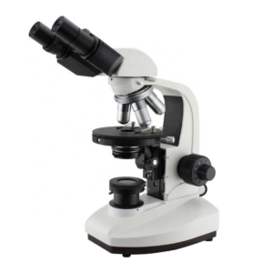
$2,090.00
The Trinocular Mercury Lamp Fluorescence Microscope utilizes the principle of fluorescence to illuminate the object by using a mercury lamp as a light source. The mercury lamp gives accurate, fast, and reliable results due to its ability to produce bright spectral lines within the visible region of the UV spectrum.
The Trinocular Mercury Lamp Fluorescence Microscope is equipped with two eyepieces (binocular) and an additional third eyepiece tube to connect a microscope camera, making it trinocular. It has a focus system consisting of fine and coarse adjustment knobs. It also contains a quadruple-reversed nosepiece, double-layer mechanical working stage, abbe condenser, collector for placing a halogen lamp, and blue and frosted filters. The light source is provided by a mercury lamp and a halogen lamp.
ConductScience offers the Trinocular Mercury Lamp Fluorescence Microscope.

Experts in Manufacturing and Exporting Optical Instruments, Microscopes and more other Products.


| Eyepiece | WF 10X (Φ18mm) |
| Eyepiece Tube | Trinocular, inclined 30° |
| Objective | Plan Achromatic Objectives PL 4X/0.10 Plan Achromatic Objectives PL 10X/0.25 Plan Achromatic Objectives PL 20X/0.40 Plan Achromatic Objectives PL 40X/0.65 (Spring) Plan Achromatic Objectives PL 100X/1.25 (Spring, Oil) Fluorescence Objective FL 25X /0.65 Fluorescence Objective FL 40X /1.00 (Spring, Glycerin) |
| Focus System | Coaxial coarse/fine focus system, with tension adjustable and up stop, minimum division of fine focusing is 2.0μm |
| Nosepiece | Quadruple (Backward ball bearing inner locating) |
| Working Stage | Double-layer mechanical (Size:160mmX140mm, moving range:75mmX50mm) |
| Abbe Condenser | N.A.1.25 Rack & pinion adjustable |
| Filter | Blue filter Froster filter |
| Collector | For Halogen Lamp |
| Light Source | 6V 20W Halogen Lamp, adjustable brightness |
| Reflected Fluorescence System | Mercury Lamp House 100W/DC Power Supply Unit: AV: 110V or 220V Fluorescence filter system B Exciton Wavelength: 450~490nm Fluorescence filter system G Exciton Wavelength: 495~555nm |
The Trinocular Mercury Lamp Fluorescence Microscope utilizes the principle of fluorescence to illuminate the object by using a mercury lamp as a light source. The mercury lamp gives accurate, fast, and reliable results due to its ability to produce bright spectral lines within the visible region of the UV spectrum.
It has two eyepieces similar to traditional binocular microscopes and contains an additional eyetube for connecting a microscope camera. Which not only captures the real-time images but also records videos. The recorded observations can be saved for future use, allowing detailed and prolonged analysis.
The Trinocular Fluorescence Microscope is highly precise and produces clear, fine images with the slightest division of 2 microns. It also includes a 6V 20W halogen lamp for transmission illumination.
The Trinocular Mercury Lamp Fluorescence Microscope is equipped with two eyepieces (binocular) and an additional third eyepiece tube to connect a microscope camera, making it trinocular. It has a focus system consisting of fine and coarse adjustment knobs. It also contains a quadruple-reversed nosepiece, double-layer mechanical working stage, abbe condenser, collector for placing a halogen lamp, and blue and frosted filters. The light source is provided by a mercury lamp and a halogen lamp. The fluorescence system is composed of a filter system for B Exciton Wavelength: 450~490nm and a filter system for G Exciton Wavelength: 495~555nm.
It works on the principle of fluorescence that illuminates the sample through excitation and emission wavelengths from the microscope’s light sources. The microscope’s camera captures the images of the specimen or records videos.
The Trinocular Mercury Lamp Fluorescence Microscope is used in various fields to identify and observe specimens. For example, it is used to identify cells, infectious agents, toxins, bioaerosol, suspicious munitions, tumors, microbial aerosols, microscopic organisms, and water quality. Its diverse usage credit goes to the microscope’s mercury lamp which has a wide and visible spectral band range.
The Trinocular Mercury Lamp Fluorescence Microscope can effectively diagnose various conditions. It can identify soluble toxins and viruses with fluorescent antibodies that are impossible to detect with other microscopes. Sanborn, Heuck, & Aouad (n.d.) used a fluorescence microscope to demonstrate staphylococcal enterotoxin infection in an animal’s body and localized its activity in the brain and other organs where it produced toxic symptoms.
It is often used in public health labs to test oil, water, and air as it has the sensitivity to detect even dead or impaired microorganisms.
The precise location, development, and function of tissues and cells can be detected using fluorochromes and fluorescent antibodies. Subcellular components such as cell wall structure, composition, and nucleic acids may also be marked by fluorochromes, depending on characteristics such as the specimen’s chemical composition, viscosity, and pH.
The Trinocular Mercury Lamp Fluorescence Microscope comes with a high-resolution camera that provides real-time specimen observation, saving time, as there are no risks of wrong observations. Bright and digitally controlled LED light sources and straightforward software enable the production of publication-quality photographs in just a few clicks. It has powerful imaging features to handle time-lapse movies, multiwell plate scanning, picture stitching and tiling, and cell counting. The microscope minimizes the complexity of high-end microscopy without losing performance.
| collector | |
|---|---|
| eyepiece | |
| filter | |
| objective | Fluorescence Objective FL 25X /0.65, Fluorescence Objective FL 40X /1.00 (Spring, Glycerin), Plan Achromatic Objectives PL 100X/1.25 (Spring, Oil), Plan Achromatic Objectives PL 10X/0.25, Plan Achromatic Objectives PL 20X/0.40, Plan Achromatic Objectives PL 40X/0.65 (Spring), Plan Achromatic Objectives PL 4X/0.10 |
| Working Voltage | American Standard: ~110V/60HZ ~230V/50HZ |
You must be logged in to post a review.
There are no questions yet. Be the first to ask a question about this product.
Monday – Friday
9 AM – 5 PM EST
DISCLAIMER: ConductScience and affiliate products are NOT designed for human consumption, testing, or clinical utilization. They are designed for pre-clinical utilization only. Customers purchasing apparatus for the purposes of scientific research or veterinary care affirm adherence to applicable regulatory bodies for the country in which their research or care is conducted.
Reviews
There are no reviews yet.