Inverted Microscope
Inverted optical microscope with objective lenses positioned below the specimen stage for examination of samples in culture vessels and larger containers.

Louise Corscadden, PhD
Director of Science · ConductScience
Ask Louise about Inverted Microscope fit, setup, configuration, or quote prep.
Already working with us? Sign in to connect this with My Scientist.
Key Specifications
Full details →- Model fit
- C. elegans, Drosophila, Gerbil, Hamster, Rabbit, Mouse, Rat, Zebrafish, Guinea pig
- SKU family
- BIO-0541
- Sizing
- 42.0 x 43.6 x 38.0 cm
- Ordering
- Online checkout and quote request available
- Category
- Microscopy
- Build notes
- Confirm accessories, station layout, and support needs before purchase
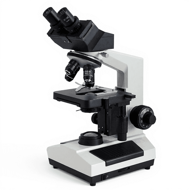
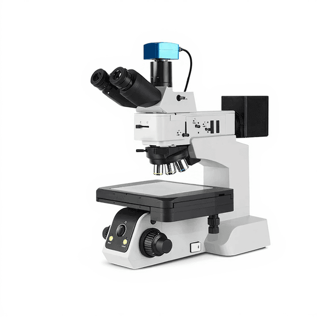
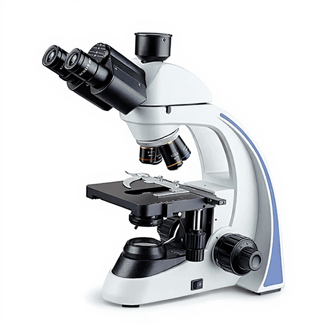
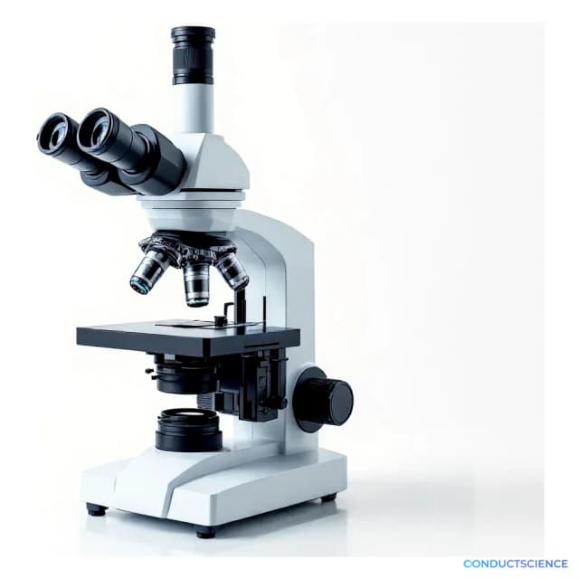
The Inverted Microscope (BIO-0541) provides essential optical magnification capabilities for biological research applications. This instrument features an inverted optical design where the objective lenses are positioned below the specimen stage, allowing observation of samples from underneath. The configuration enables examination of specimens in culture vessels, petri dishes, and other containers that cannot be easily inverted.
The inverted design accommodates larger specimens and culture vessels while maintaining stable optical performance. Researchers utilize this microscope for routine cell culture monitoring, basic histological examinations, and general biological sample observation where standard upright microscopy is impractical due to sample container geometry or size constraints.
How It Works
The inverted microscope operates on standard light microscopy principles with the optical components arranged in reverse orientation compared to conventional upright systems. Illumination originates from above the specimen stage, passing through the sample and into objective lenses positioned below. This configuration places the light source and condenser system above the specimen, while the objective turret and eyepieces are located beneath the stage.
Light transmitted through the specimen is collected by the inverted objective lenses and directed through the optical tube system to the eyepieces or camera port. The inverted design eliminates working distance limitations imposed by culture vessel walls and allows examination of samples in their natural growth orientation. The stage assembly provides mechanical stability while accommodating various container sizes and shapes that would be incompatible with upright microscope geometry.
Features & Benefits
Automation Level
- manual
Research Domain
- Analytical Chemistry
- Cancer Research
- Cell Biology
- Developmental Biology
- Histopathology
- Microbiology
Species
- C. elegans
- Drosophila
- Gerbil
- Hamster
- Rabbit
- Mouse
- Rat
- Zebrafish
- Guinea pig
Weight
- 15.0 kg
Dimensions
- L: 42.0 mm
- W: 43.6 mm
- H: 38.0 mm
| Feature | This Product | Typical Alternative | Advantage |
|---|---|---|---|
| Optical Design | Inverted configuration with bottom-mounted objectives | Entry-level models may offer only upright configurations | Enables examination of specimens in culture vessels without sample transfer or inversion requirements. |
| Base Construction | 15 kg stable base construction | Lighter models may offer reduced stability | Provides mechanical stability for extended observations and reduces vibration artifacts during critical measurements. |
| Stage Accessibility | Large stage area accommodating various vessel sizes | Compact models may have limited stage dimensions | Supports diverse culture vessel types and sizes common in cell biology and microbiology applications. |
| Illumination System | Transmitted light illumination from above | Basic models may have limited illumination control | Maintains consistent lighting conditions for various sample types and container materials. |
This inverted microscope provides essential capabilities for culture vessel examination with stable mechanical construction and standard optical performance. The design accommodates various sample container types while maintaining focus stability during extended observation periods.
Practical Tips
Verify objective parfocality using a test specimen when first installing or changing objective lenses.
Why: Proper parfocality ensures minimal refocusing when changing magnifications, improving workflow efficiency.
Clean objective lenses weekly or after each use with lens paper and appropriate cleaning solution, working from center outward.
Why: Oil, dust, and sample residue on objectives significantly degrade image quality and contrast.
Allow culture vessels to reach room temperature before observation to minimize condensation and thermal drift.
Why: Temperature differences cause focus drift and condensation that obscures sample details during observation.
Check condenser alignment and lamp centering if illumination appears uneven across the field of view.
Why: Uneven illumination creates measurement artifacts and makes comparative observations unreliable.
Use consistent illumination intensity settings when comparing samples or documenting results across different sessions.
Why: Variable lighting conditions affect image contrast and can introduce measurement inconsistencies.
Ensure culture vessels are properly sealed and handle biological samples according to laboratory safety protocols.
Why: Inverted microscopy may require longer observation times, increasing exposure risk with biological specimens.
Position the microscope away from air conditioning vents and high-traffic areas to minimize vibration and temperature fluctuations.
Why: Environmental stability directly impacts focus stability and image quality during extended observations.
Setup Guide
What’s in the Box
- Inverted microscope main unit (typical)
- 10x eyepieces (typical)
- 10x and 40x objective lenses (typical)
- Power adapter and cord (typical)
- Stage plate assembly (typical)
- User manual and documentation (typical)
- Dust cover (typical)
Warranty
ConductScience provides a standard one-year manufacturer warranty covering defects in materials and workmanship, with technical support available for setup and operational questions.
Compliance
References
Background reading relevant to this product:
What types of culture vessels can be accommodated with the inverted design?
The inverted configuration accommodates standard tissue culture flasks, multi-well plates, petri dishes, and other containers up to the stage size limitations. The bottom-mounted objectives eliminate height restrictions common with upright systems.
How does the optical performance compare to upright microscopes?
Optical performance follows standard microscopy principles with comparable resolution and image quality. The inverted design may introduce slight differences in working distance and condenser positioning but maintains equivalent magnification and clarity capabilities.
What maintenance procedures are required for the optical components?
Regular cleaning of objective lenses, eyepieces, and condenser elements using appropriate optical cleaning solutions. Dust covers should be used when not in operation, and mechanical components require periodic lubrication according to manufacturer specifications.
Can the microscope be upgraded with camera systems for documentation?
Most inverted microscopes include camera ports or can be adapted for digital imaging systems. Verify camera mount compatibility and optical tube specifications for your specific documentation requirements.
What illumination options are compatible with this system?
Standard transmitted light illumination is provided with potential for phase contrast, differential interference contrast, or fluorescence modules depending on the specific model configuration and available accessories.
How stable is the focus during extended observation periods?
Mechanical stability depends on the base construction and environmental conditions. Temperature fluctuations and vibrations can affect focus stability, requiring periodic readjustment during long observation sessions.
What sample thickness limitations exist with the inverted design?
Sample thickness is limited by the working distance of the objective lenses and the depth of the culture vessel or slide. Consult objective specifications for maximum working distances with your sample types.
Have a question about this product?
Have a question? Just ask.
Send it over and we'll email you a personalized answer — no call, no scheduling.
Prefer to talk it through?
Accessories
Enhance your setup with compatible accessories
Frequently Bought Together
Related Products