$7,990.00 – $8,590.00Price range: $7,990.00 through $8,590.00
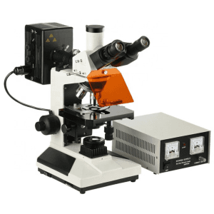
An inverted fluorescent microscope uses fluorescence to study specimens, such as living cells at the bottom of a petri dish or tissue culture. The Inverted Fluorescent Microscope has similar components to those of other microscopes, and the only difference is the arrangement of the parts, which are placed in inverted positions.
The specimen is lit using a halogen lamp as a light source. When light enters the microscope, it strikes a dichroic mirror, reflecting one range of wavelengths while allowing another to pass through. The ultraviolet light is reflected up to the specimen by the dichroic mirror. The UV light causes fluorescence in the specimen’s molecules and the fluorescent-wavelength light generated is collected by the objective lens.
ConductScience offers the Inverted Fluorescent Microscope.

Conduct Science is a premier manufacturer of research infrastructure, born from a mission to standardize the laboratory ecosystem. We combine industrial-grade precision with a scientist-led tech-transfer model, ensuring that every instrument we build solves a real-world experimental challenge. We replace "home-brew" setups with validated tools ranging from microsurgical suites to pathology systems. With a track record of >1,600 institutional partners and hundreds of citations, our equipment is engineered to minimize human error. We help you secure more data for less of your budget, delivering the reliability required for high-impact publication.

bool(false)

bool(false)

Eyepiece | WF 10X (Φ20mm) |
|---|---|
| Eyepiece Tube | Trinocular, inclined 30° (100% image light for photography capable) |
| Objective | Long working distance plan achromatic objectives PL L10X/0.25 Long working distance plan achromatic objectives PL L25X/0.40 Long working distance plan achromatic objectives PL L40X/0.60 Long working distance plan achromatic phase contrast objective PL L10X/0.25 PHP2 Long working distance plan achromatic phase contrast objective PL L25X/0.40 PHP2 Long working distance plan achromatic phase contrast objective PL L40X/0.60 PHP2 |
| Focus System | Coaxial coarse/fine focus system, with tension adjustable and up stop, minimum division of fine focusing is 2.0μm |
| Nosepiece | Quintuple (Ball bearing inner locating) |
| Working Stage | Double-layer mechanical (Size:224mmX208mm,moving range:112mmX79mm) |
| Filter | Blue Filter Green Filter |
| Shelf of specimen | Match to install 68mm or 77mmX29mm culture utensil Match to install 82mmX57mm culture utensil Match to install 128mmX85mm 96 holes culture utensil |
| Reflected fluorescence system | Mercury lamp house 100W/DC Power supply unit: AV:110V or 220V Fluorescence filter system B exciton wavelength:450~490nm Fluorescence filter system G exciton wavelength:495~555nm Fluorescence filter system UV exciton wavelength:320~380nm Fluorescence filter system V exciton wavelength:380~415nm |
| Turnplate phase contrast unit | Centering Telescope Condenser Rack & Pinion adjustable, NA=0.4, working distance: 50mm |
An inverted fluorescent microscope uses fluorescence to study specimens, such as living cells at the bottom of a petri dish or tissue culture. It is called “inverted” due to the arrangement of the microscope’s components, which are placed in inverted positions. The objective lenses are located above the stage, while the condenser and the light source are below the stage. The primary purpose of the condenser lens above the specimen stage is to focus light on the specimen. Conduct Science’s Inverted Fluorescent Microscope is equipped with wide-field eyepieces that allow images to be obtained in a wide view field. It also contains plain achromatic objectives that allow a longer working distance of 50 mm.
Fluorescence microscopy utilizes fluorescent dyes, called fluorophores, that absorb one wavelength of light (the excitation wavelength) and emit the second, longer wavelength (the emission wavelength). Since most cell molecules are not fluorescent, the experimenter introduces fluorescent labels to be imaged. Therefore, the labels can be targeted to the desired molecules by attaching fluorescently labeled antibodies or genetically encoding a fluorescent protein. This allows the identification of different fluorescent molecules at the same time, which can be detected at extremely low abundance, making it a very powerful technique (Thorn, 2016).
The microscope’s halogen lamp serves as excitation light and illuminates the sample through the same objective that detects the light emission. Interference from the excitation light is eliminated through a fluorescence filter cube that separates light by wavelength, allowing the emitted light to be imaged.
The Inverted Fluorescent Microscope has similar components to those of other microscopes, and the only difference is the arrangement of the parts, which are placed in inverted positions. Apart from its similar components such as the stage, objective lens, eyepiece, eyepiece tube, dual-concentric knobs, nosepieces, and condenser lens, it also consists of other elements. A 6V 30W Halogen lamp is present with a brightness control that serves as an excitation lamp that illuminates the sample. It contains an excitation filter that filters all wavelengths of the light source except for the excitation range of the fluorophore under investigation. It also contains an emission filter that filters out the excitation range and transmits the emission range of the fluorophore under investigation. A dichroic mirror is placed between the two filters that serve as a beam splitter.
The specimen is lit using a halogen lamp as a light source. When light enters the microscope, it strikes a dichroic mirror, reflecting one range of wavelengths while allowing another to pass through. The ultraviolet light is reflected up to the specimen by the dichroic mirror. The UV light causes fluorescence in the specimen’s molecules and the fluorescent-wavelength light generated is collected by the objective lens. The fluorescent light then goes via a dichroic mirror and the emission filter before reaching the eyepiece and forming the picture of the targeted specimen (Sanderson, Smith, Parker, & Bootman, 2014).
The Inverted Fluorescent Microscope can be used to observe and detect various living cells and microscopic organisms at the bottom of lab vessels like tissue culture flasks and Petri dishes. For example, it can be used to identify Phytophthora spp, nematology extraction specimens such as vermiform, and to see Mycobacterium tuberculosis. Moreover, micromanipulation is also possible with this microscope
Wei et al. (2019) developed a nano-drug of doxorubicin and exosome derived from mesenchymal stem cells to treat osteosarcoma in vitro. They used an inverted fluorescent microscope and flow cytometry to analyze the cellular absorption of Exo-Dox and free Dox by myocardial H9C2 cells since the major side effect induced by doxorubicin is cardiac toxicity. An exosome isolation kit was used to separate the exosome from bone marrow. The exosome and Exo-Dox were characterized using nanoparticle tracking analysis and a transmission electron microscope.
In a study conducted by Sun et al. (2019), they used an Inverted fluorescent microscope and a hyperspectral microscope to explain the exact mechanism of how Escherichia coli O157:H7 interacts and colonizes fresh cucumber (Cucumis sativus L.). They found out that E. coli O157:H7 occupied the epidermis of cucumbers largely around the stomata without penetrating the interior tissues. After infiltrating the internal tissues, the bacteria colonized the cucumber placenta and vascular bundles (particularly the xylem vessels) and migrated along the xylem vessels. Furthermore, E. coli O157:H7 moved quicker from the stalk to the flower bud than from the flower bud to the branch.
The inverted fluorescent microscope has a large stage to see specimens in glass tubes and Petri plates. Its design allows the observation and analysis of cells in vast quantities of media and cell tissues in their native vessel, bigger than microscopic slides, making it superior to the upright microscope. It has three different specimen holders. It maintains the sterility of the sample since the specimen is not contaminated by contact with the objective lens. The microscope can be used with differential interference contrast and phase-contrast optics to improve picture quality. It allows the connection of a wide range of digital recording devices. The wide-field eyepieces give clear images of up to 2-micron fine focusing, and a large view field is obtained using simple, achromatic objectives with a long working distance (no cover glass).
Photobleaching occurs when fluorophores lose their capacity to glow when they are illuminated. Photobleaching reduces the time a sample can be viewed using fluorescence microscopy; it may be reduced by various methods, including the use of more robust fluorophores, reduced light, and the application of photoprotective scavenger chemicals.
Turn off the power during installation, assembly, cable connection/disconnection, and maintenance.
Wet or spilled substances on the device might cause it to malfunction, overheat, or electrocute you.
Hold the product firmly by the bottom front recess and the bottom rear recess while carrying it.
Prevent scratches on optical components such as lenses and filters because scratches can diminish the focus of the microscopic image.
Dispose of contaminated slides according to your facility’s regular procedures to eliminate biohazard concerns.
Place the microscope in an area with a temperature range of 0 to 40°C with a relative humidity of 85% or less.
Avoid physical shocks and vibrations while handling the goods. Even little physical shocks have the potential to degrade the accuracy of targets.
Wei, H., Chen, J., Wang, S., Fu, F., Zhu, X., Wu, C., Liu, Z., Zhong, G., & Lin, J. (2019). A Nanodrug Consisting Of Doxorubicin And Exosome Derived From Mesenchymal Stem Cells For Osteosarcoma Treatment In Vitro. International journal of nanomedicine, 14, 8603–8610. https://doi.org/10.2147/IJN.S218988
Sun, Y., Wang, D., Ma, Y., Guan, H., Liang, H., & Zhao, X. (2019). Elucidating Escherichia Coli O157:H7 Colonization and Internalization in Cucumbers Using an Inverted Fluorescence Microscope and Hyperspectral Microscopy. Microorganisms, 7(11), 499. https://doi.org/10.3390/microorganisms7110499
Sanderson, M. J., Smith, I., Parker, I., & Bootman, M. D. (2014). Fluorescence Microscopy. Cold Spring Harbor Protocols, 2014(10), pdb.top071795. https://doi.org/10.1101/pdb.top071795
Thorn, K. (2016). A quick guide to light microscopy in cell biology. Molecular Biology of the Cell, 27(2), 219–222. https://doi.org/10.1091/mbc.e15-02-0088
| Weight | 110.23 lbs |
|---|---|
| Dimensions | 75 × 55 × 35 cm |
| model | MCX-VMF200I, MCX-VMF400I |
| Brand | ConductScience |
You must be logged in to post a review.
There are no questions yet. Be the first to ask a question about this product.
Reviews
There are no reviews yet.