Introduction
Endotracheal tubes are used for the maintenance and establishment of clear airways in subjects. These tubes reduce anatomical dead space and allow an easy performance of assisted or mechanical breathing procedures when required. These specific tracheal tubes are almost always inserted through the mouth (orotracheal) or the nose (nasotracheal) into the trachea. The size of the endotracheal tubes is dependent on the size and weight of the subject, and hence, they vary for every species and individual.
Apparatus And Equipment
Endotracheal tubes can be made of PVC, polyethylene, polyurethane, silicone rubber, and red rubber. Often reusable tubes are made of rubber. However, with time they tend to deteriorate, become brittle, and are easily kinked. The use of red rubber has also decreased due to its opaque nature and latex allergies.
PVC, polyethylene, and polyurethane have many advantages over rubber tubes. The clear tubes show the appearance of condensation with each breath when placed correctly. On the other hand, silicone tubes are translucent and tend to be relatively resistant to kinking. In certain situations where there is excessive flexing of the head and neck of the subject, using an armored tube reinforced with a wire coil can prevent the tube from kinking while still being flexible enough. However, these tube types should not be used during the MRI procedure.
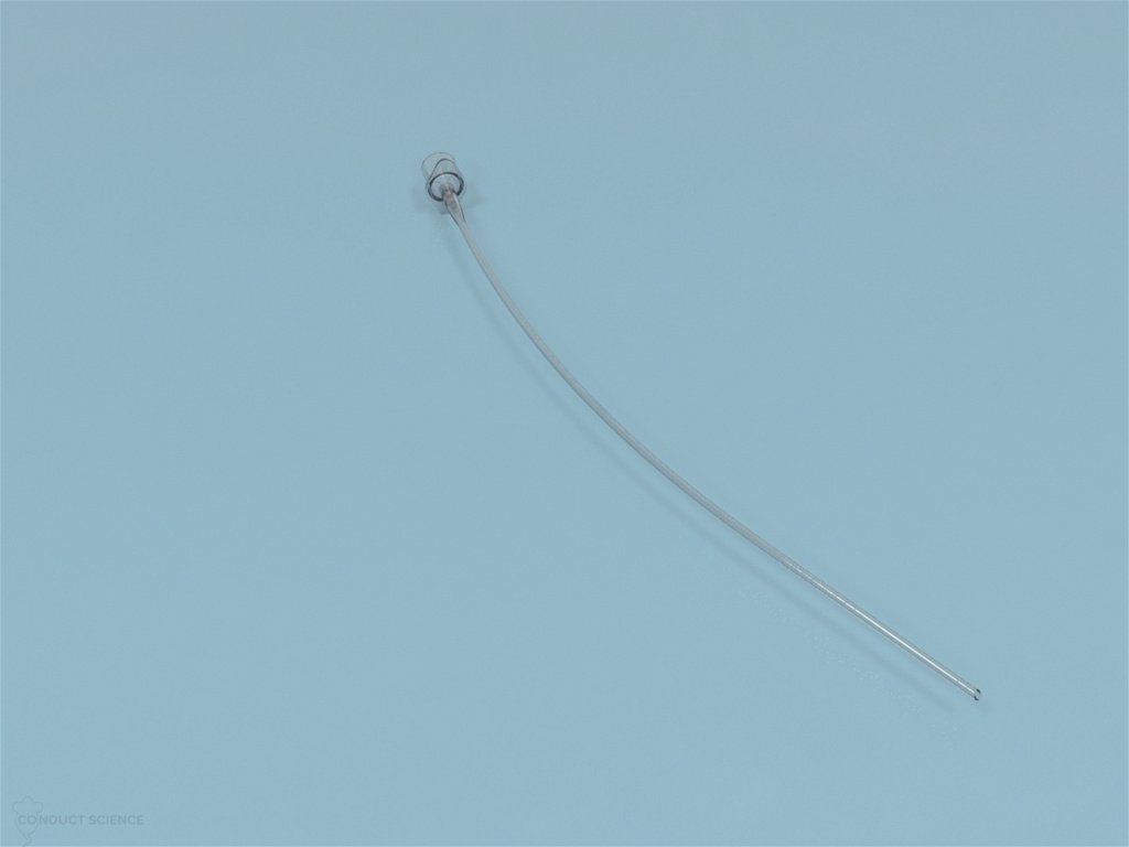
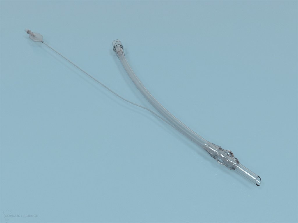
Endotracheal tubes usually will have a preformed curve to allow better visualization of laryngeal opening during intubation. The tubes have a beveled distal end and may also have a small opening opposite the bevel called the Murphy eye as an additional path for gas in case the bevel is occluded.
Types Of Endotracheal Tubes
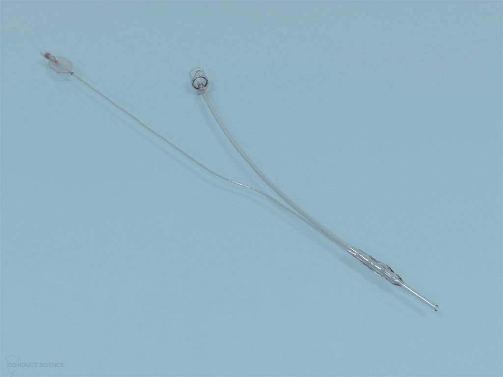
Endotracheal tubes are primarily either cuffed or uncuffed. The cuffed tubes provide a seal around the tube to prevent leakage of anesthetic gases and aspiration of oral secretions or gastric contents. The cuffed endotracheal tubes are available with cuffs that are low-volume, high-pressure cuffs or high-volume, low-pressure cuffs. Low-volume, high-pressure cuffs provide a narrow seal at the tracheal wall and require high pressure for inflation, while the latter type of cuff allows a greater area of tracheal contact and requires low inflation pressure.
The cuff is inflated via a channel in the tube, equipped with an inflation line, using either a pilot balloon or a 2 to 5 ml syringe. Usually, gases such as nitrous oxide are used to inflate the cuff, but in the case of silicone tubes using the saline solution is recommended due to its permeability to gases.
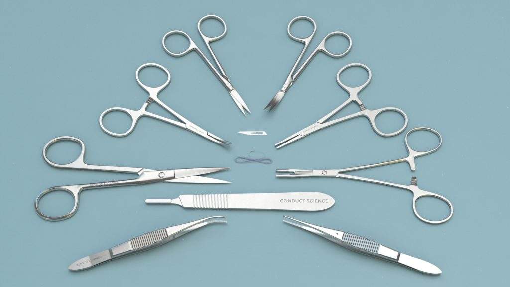
The deflation of the cuff can be prevented by the non-return valve often present on most disposable tubes or by clamping using a pair of hemostats.
Mode Of Operation
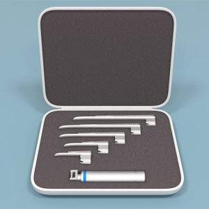
Endotracheal tubes are selected based on the species and the size of the subject. A corresponding laryngoscope is also selected based on the same parameters. Commercially available endotracheal tubes tend to be excessively long. Thus, it must be cut down to an appropriate length usually to the approximate distance from the external nares to just anterior to the thoracic inlet. For small animals, an uncuffed endotracheal tube may be a better option. Lubrication of the tubes with a small quantity of lidocaine gel will allow easy passage of the tubes.
Before intubation, the subject must be adequately anesthetized to prevent cough and swallowing reflexes. Also, subjects should be administered with oxygen for about 2 minutes before being intubated to slow the advent of hypoxia should the larynx be obstructed.
Endotracheal Intubation Methodology
Cat, Dog, and Sheep
Place the subject in sternal recumbency, opening its jaw as wide as possible. Drawing the subject’s tongue forward, advance the laryngoscope towards the pharynx. Apply gentle upward pressure on the subject’s soft palate using the end of the endotracheal tube to disengage the epiglottis to gain an obstructed via of the larynx. It is advisable to spray the larynx of cats and sheep to prevent laryngospasm. Advance the endotracheal tube through the larynx into the trachea and connect the tube to the anesthetic breathing system. If using a cuffed tube, inflate it appropriately, and tie the tube to the animal’s jaw to prevent displacement. Perform assisted ventilation and observe the movement of both sides of the thorax to ensure correct positioning of the tube. Additionally, manual inflation can be performed to observe any issues with the endotracheal tube and its placement.
Pig
Place the subject on its back or chest and extend the tongue taking care not to damage its surface on the teeth. Place an introducer inside the tube to straighten and ease the process of tube advancement.
Advance the laryngoscope over the tongue and disengage the epiglottis by pushing on the soft palate using the tip of the introducer if necessary. Spray the larynx with lidocaine once the larynx is visible. Gently advance the introducer-endotracheal tube into the larynx and withdraw the introducer. Gently advance the tube further. If resistance is felt, withdraw the tube slightly, rotate 90°, and reinsert. Repeat this process until no resistance is experienced. Avoid forcing the tube through the larynx as this can result in severe trauma, edema, hemorrhage, and consequent asphyxiation.
Rat
Place the subject on its back and gently extend its tongue forward and to one side. Advance the purpose-made laryngoscope or otoscope until the larynx is visualized. Intubation in rats can be performed using arterial cannula between sizes 12 to 16 gauge that has a rubber tubing or certain micropore tape positioned at about 0.75 to 1 cm from the tip to serve as the cuff. Connect the tube to the anesthetic machine using a modified Luer fitting ensuring absolute minimum dead space. Using a silk ligature glued to the base of the Luer mount, anchor the catheter to the subject’s jaw. If using an otoscope, make use of an introducer to pass the cannula.
Birds
Open the subject’s beak and gently pull the tongue forward. For larger birds, standard pediatric tubes (>2.5 mm) can be used, while in smaller birds it is advisable to use intravenous or urinary catheters cut to an appropriate length. In birds, only uncuffed tubes should be used since cuffed tubes can cause pressure necrosis of tracheal mucosa.
Rabbit
Place the subject on its back and gently pull the tongue forwards taking care to avoid contact with sharp edges of incisors. Introduce the otoscope or laryngoscope and advance it until the larynx is visible. In case the paler triangle of the epiglottis is seen, the structure can be manipulated to allow a clear view of the larynx. The larynx can be sprayed with lidocaine if necessary. Pass the purpose-made introducer (or a dog/cat urinary catheter without Luer fitting) through the otoscope into the larynx and the trachea. After placement of the introducer, remove the otoscope or laryngoscope, ensuring the position of the introducer is not changed. Thread the endotracheal tube onto the end of the introducer. If resistance is felt during the advancement of the endotracheal tube, gently rotate the tube to ease the process. Remove the introducer only after the tube is successfully placed in the trachea. Tie the tube in place to avoid displacement.
Alternatively, blind intubation can be performed on the subject. Place the subject in sternal recumbency position. Gripping the head firmly and extended, lift the subject such that its forelegs are just touching the operating table. Advance the endotracheal tube over the tongue through the gap between the incisors and the premolars towards the larynx. Listen for breath sounds or in the case of a clear tube, observe for condensation. Gently advance the tube, giving a quarter turn as it enters the larynx to ease the passage.
Intranasal Intubation
Intranasal intubation is often used as an alternative to face mask. Lubricate the tube before insertion. Generally, the tube should be advanced medially and ventrally. Rotate the catheter slightly to aid insertion. A Luer adapter and oxygen bubble tubing can be used to connect the catheter to the anesthetic machine.
Precautions
For cuffed tubes, the cuff must be inflated before use to inspect for tears and evenness of inflation. Wash the tubes thoroughly using hot soapy water or pasteurize when possible. Few endotracheal tubes may be autoclaved or sterilized using ethylene oxide. If using the latter method for sterilization, eliminate all traces of the gas before use. Disposable endotracheal tubes are preferred to minimize the risk of infection.
References
- Fish, R. E. (2008). Anesthesia and analgesia in laboratory animals. Amsterdam: Elsevier.
- Flecknel, P. (2009). Laboratory Animal Anaesthesia. Elsevier.


































Reviews
There are no reviews yet.