x
[quotes_form]

Stereotaxic

Stereotaxic Surgery Innovation

Featured Product

Stereotaxic Precise Spinal And Cortical Impactor

The Conduct Science precise impactor is used to create and evaluate traumatic brain injury mechanisms in pre-clinical medical research.

This device allows researchers to induce traumatic brain injuries (TBIs) with parameters such as impact depth, velocity, and dwell time precisely controlled to ensure accurate damage to provide high efficiency, repeatability, and stability, ensuring the reproducibility ofĀ  impact direction and injury position.

Moreover, the precise impactor assesses cord injury mechanisms effectively. It can be precisely locked and applied to the spinal cord, especially when combined with our spinal cord adapters designed for mice and rats.

We understand that research and clinical needs can be unique and specific. Therefore, we offer custom solutions tailored to meet specific research and clinical requirements. Our custom solutions include:

  • Customized Frames and Components: Designed to fit unique experimental setups and specifications.
  • Bespoke Systems: Tailored instruments and setups to meet specific research and clinical requirements.
  • Collaborative Design Process: We work closely with researchers and clinicians to develop solutions that address their precise needs.
  • Comprehensive Support: Includes installation, training, and ongoing technical support to ensure successful implementation and us

Overview

Stereotaxic instruments are essential tools in neurosurgery and medical research, enabling precise targeting within the brain and other parts of the body. These instruments employ a three-dimensional coordinate system to locate specific areas, facilitating minimally invasive procedures such as injections, stimulations, biopsies, and radiosurgery. By accurately pinpointing the affected areas, stereotaxic instruments help avoid damage to surrounding healthy tissues, making surgeries safer and more effective. This precision is particularly valuable in treating small tumors, which can be sampled, tested, and irradiated with minimal impact on adjacent areas. Stereotaxic techniques are also utilized in various experimental and therapeutic procedures, advancing both medical science and patient care.

Stereotaxic Manual

Our standard stereotaxic frames are ideal for basic surgical procedures and research applications. These frames provide a stable and reliable platform for positioning and securing subjects during procedures. Key features include:

  • High Precision: Ensures accurate targeting and reproducibility.
  • Versatility: Suitable for various experimental setups.
  • Ease of Use: Simple to set up and operate, reducing training time.
  • Durability: Constructed from high-quality materials to withstand repeated use.

Stereotaxic Manual Device

Manual Stereotaxic frame for mouse and rat with single of dual arms.

Designed for maximum precision and accuracy, the Conduct Science Stereotaxic frame featuresĀ a three-dimensional operating arm (X, Y, and Z axis) to provide aĀ reading accuracy ofĀ  0.1mm (100Ī¼m) and travel range ofĀ 80mm. To ensure optimum long-term use, the operating arm replaces the traditional metal-to-metal ā€œhard grindingā€ transmission with a combination of screw rod and rolling mechanisms to maintain the stability and accuracy of the coordinate axis over time.Ā Molded transmission screws and thread sleeves and the use of connecting joints (rather than multiple joint connections) ensure stability and uniformity to reduce error in use.

This instrument comes with a set of mouse or rat adapters and a pair of ear bars.

Stereotaxic Digital

Our Digital Stereotaxic systems are advanced tools designed for precise and reliable stereotaxic procedures across various animal models. With state-of-the-art digital integration, these systems provide enhanced accuracy and ease of use for researchers and clinicians. Key features include:

Digital Precision: Advanced digital controls for highly accurate targeting and positioning.
User-Friendly Interface: Intuitive software interface allows for easy setup and operation.
Enhanced Data Integration: Seamlessly integrates with other digital systems for comprehensive data collection and analysis.
Safety and Stability: Built with secure restraints and ergonomic design to ensure animal welfare duringĀ procedures.

Stereotaxic Advanced

For more complex surgeries and advanced research, our advanced stereotaxic systems offer enhanced features. These systems are equipped with sophisticated technology to improve accuracy and efficiency. Key features include:

  • Automated Functions: Includes motorized drives and digital controls for precise adjustments and repeatable positioning.
  • Modular Design: Allows for customization and expansion with additional accessories and components.
  • User-Friendly Software: Provides intuitive interfaces for planning and executing complex procedures.

Stereotaxic Large Animal

Our large animal stereotaxic devices are robust systems tailored for use in larger animals, ensuring precise targeting and stability during procedures. These systems are crucial for veterinary surgeries and research involving large animal models. Key features include:

  • Robust Construction: Built to handle the size and weight of larger animals while maintaining stability.
  • Precision Targeting: Ensures accurate placement for various procedures, from neurological studies to orthopedic surgeries.
  • Adjustable Components: Designed to accommodate different species and experimental requirements.
  • Safety Features: Includes secure restraints and support to ensure the well-being of the animals during procedures.

Stereotaxic Accesories

  • Adaptor Kits: Compatible with various animal models to ensure precise positioning.
  • Ear Bars and Nose Clamps: Securely hold the subject’s head in place during procedures.
  • Microinjectors: Allow for precise delivery of substances to targeted brain regions.
  • Electrode Holders: Facilitate accurate placement of electrodes for recording and stimulation.
  • Digital Displays and Controllers: Provide real-time feedback and control over stereotaxic adjustments.
  • Heating Pads: Maintain the subject’s body temperature during prolonged procedures.

History

History of Stereotaxic Rodent Surgery

Stereotaxic rodent surgery is a crucial technique in neuroscience and biomedical research, allowing precise targeting of specific areas within the brain. The history of this method dates back to the early 20th century and has evolved significantly over the decades.

Ā 

Origins and Early Developments

The concept of stereotaxy originated in human medicine. The term “stereotaxic” comes from the Greek words “stereos” (solid) and “taxis” (arrangement), reflecting the technique’s ability to position instruments accurately in three-dimensional space. In 1906, British neurosurgeon Sir Victor Horsley and Robert Henry Clarke developed the first stereotaxic apparatus for brain surgery in humans, aiming to treat conditions like epilepsy and movement disorders (Horsley & Clarke, 1908).

Ā 

Adaptation to Rodent Models

In the 1930s and 1940s, researchers began adapting stereotaxic techniques for use in smaller animals, particularly rodents. Rodents, such as rats and mice, are widely used in biomedical research due to their genetic similarities to humans and their suitability for controlled experimental conditions. The adaptation of stereotaxic surgery for rodents allowed scientists to conduct detailed and reproducible studies on brain function, neuroanatomy, and behavior (Paxinos & Watson, 1986, Paxinos, 2015).

Ā 

Expansion and Clinical Applications

By 1947, Earnest A. Spiegel and Henry T. Wycis introduced stereotactic surgery for humans, termed “stereoencephalotomy,” using adaptations from earlier animal models (Spiegel et al., 1947). Their work with a microdrive mounted on a plaster cap allowed precise electrode placement guided by pneumoencephalography, a technique pioneered by Dandy using X-rays for brain landmark identification (Dandy, 1918).

The global adoption of stereotactic surgery in neurosurgery accelerated with innovations like Lars Leksell’s arc-centered device, which standardized the procedure and facilitated its widespread use. By 1965, an estimated 25,000 treatments had been performed worldwide, primarily for conditions like Parkinson’s disease and epilepsy (Spiegel, 1965).

Ā 

Advancements in Techniques and Technology

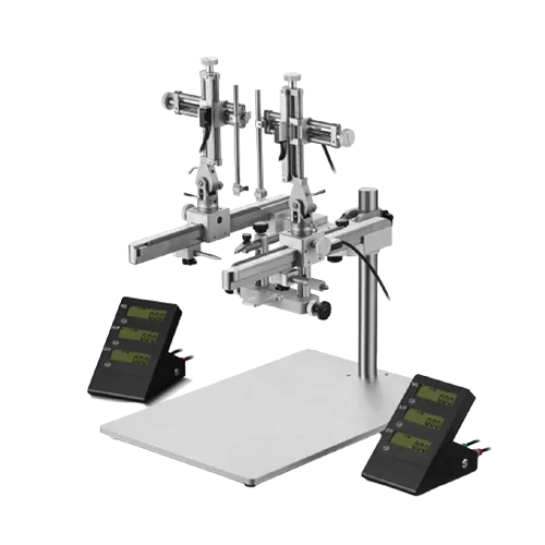
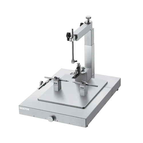
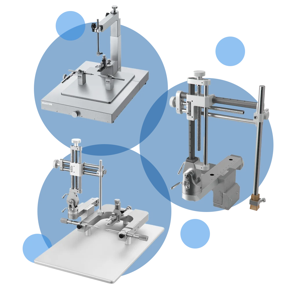
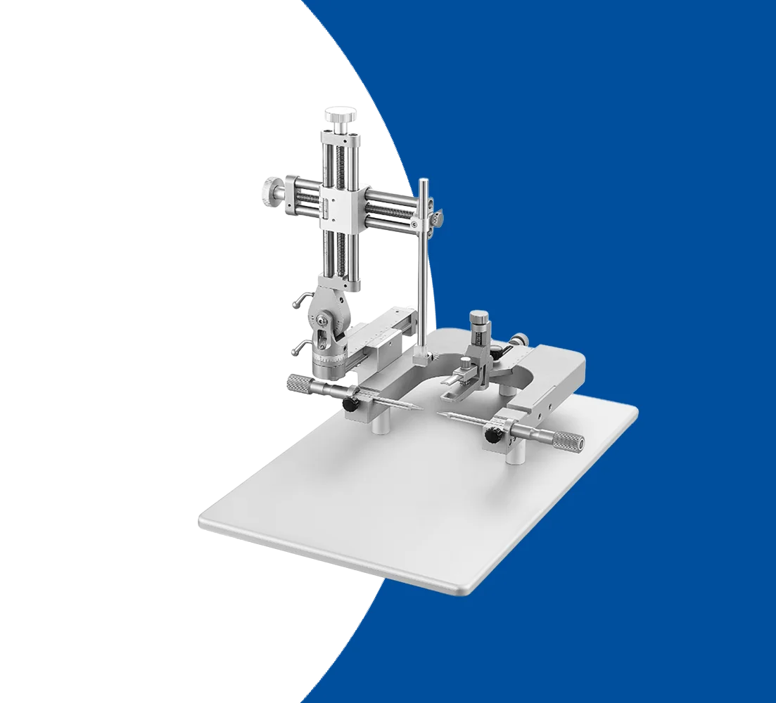
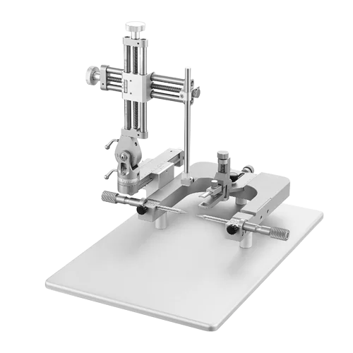
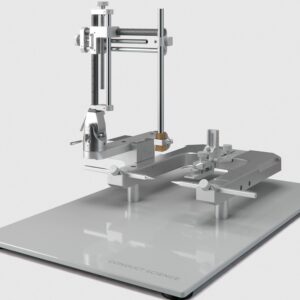
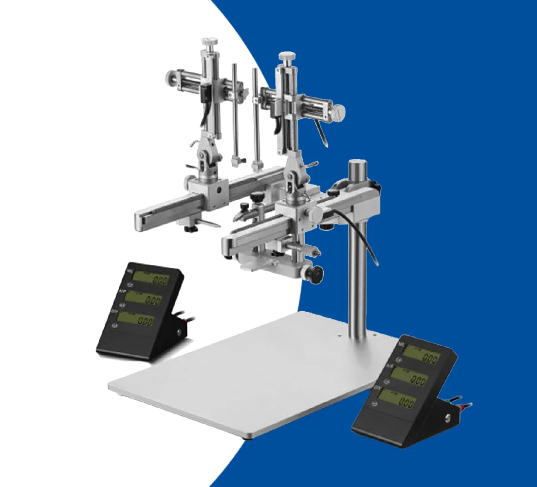
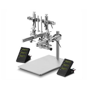
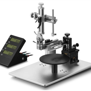
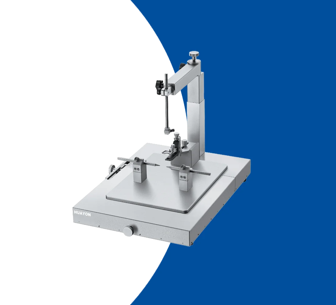
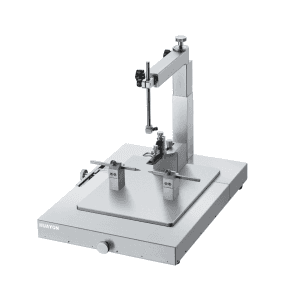
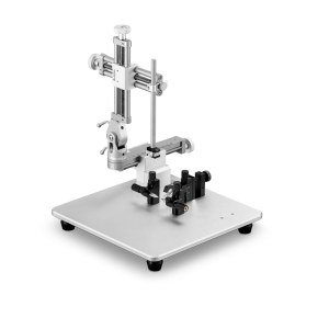
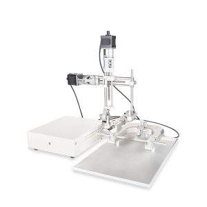
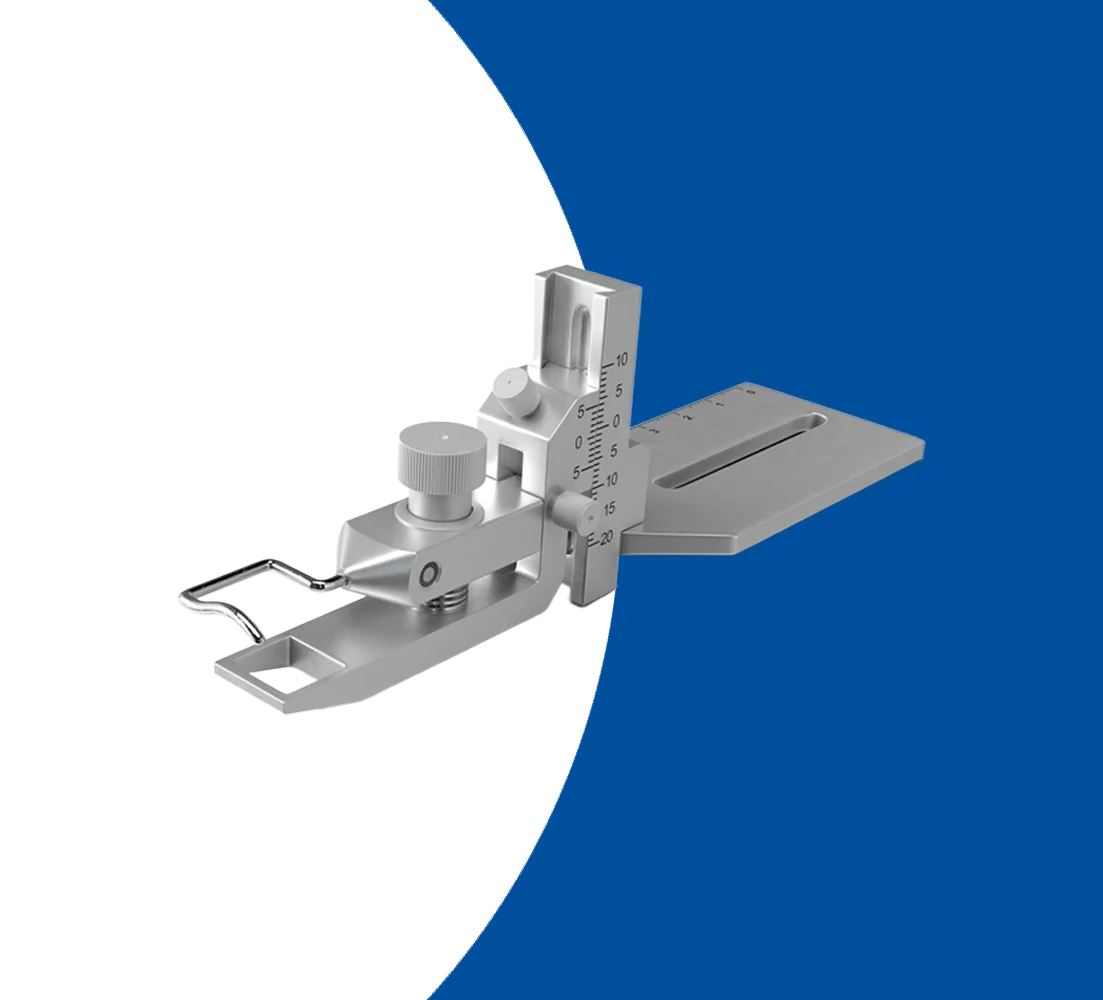
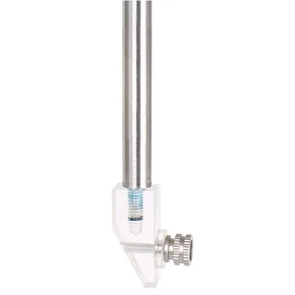
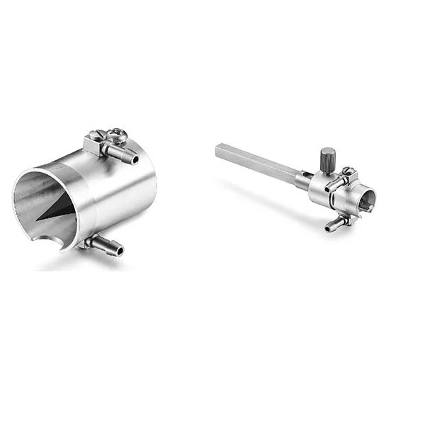
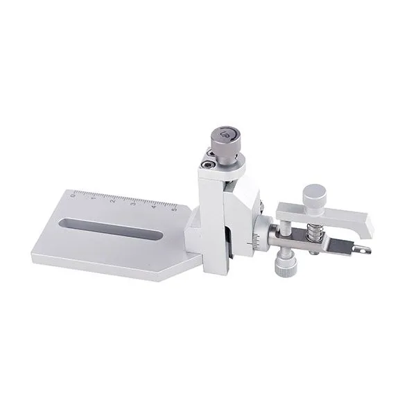
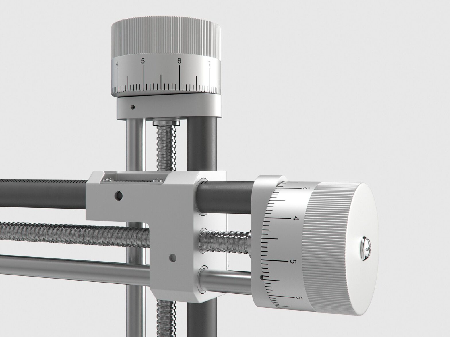
In the 1950s and 1960s, advancements in stereotaxic instruments and techniques significantly improved the precision and reliability of rodent surgeries. Innovations included more accurate atlases of rodent brain anatomy, improved surgical instruments, and the development of more sophisticated stereotaxic frames. These frames allowed for the immobilization of the animal’s head, ensuring accurate targeting of brain regions.

Ā 

Modern Applications

Today, stereotaxic rodent surgery is an essential tool in neuroscience research. It enables researchers to perform a wide range of procedures, including:

  • Lesion Studies: Creating precise lesions in specific brain regions to study their function (Paxinos, 2015).
  • Electrode Implantation: Inserting electrodes to record brain activity or deliver electrical stimulation (Swanson, 2018).
  • Drug Delivery: Administering drugs directly into specific brain areas (Wise et al., 1973).
  • Gene Manipulation: Using techniques like optogenetics and chemogenetics to control gene expression and neural activity (Yizhar et al., 2011).

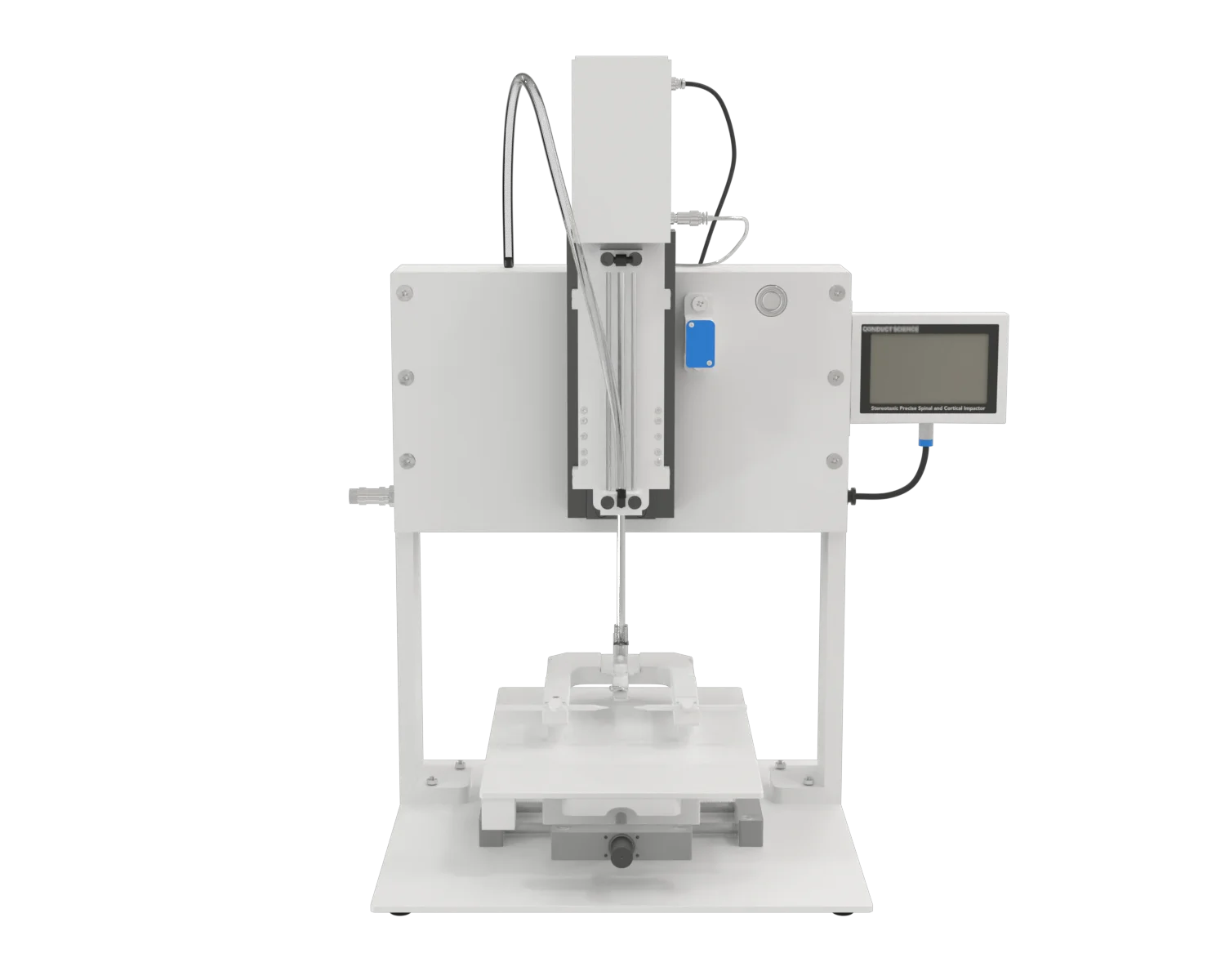
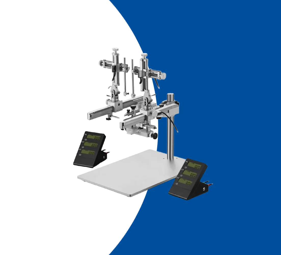
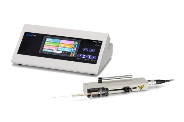
Modern stereotaxic instruments are highly sophisticated, often incorporating digital coordinates, computer-assisted guidance, and advanced imaging techniques like MRI and CT scans to enhance accuracy (Cecyn & Abrahao, 2023).

Ā 

References

Cecyn, M. N., & Abrahao, K. P. (2023). Where do you measure the Bregma for rodent stereotaxic surgery?. IBRO neuroscience reports, 15, 143ā€“148.Ā 

Dandy W. E. (1918). Ventriculography following the injection of air into the cerebral ventricles. Annals of surgery, 68(1), 5ā€“11

Horsley, V., & Clarke, R. H. (1908). The structure and functions of the cerebellum examined by a new method. Brain, 31(1), 45-124.

Paxinos, G., & Watson, C. (1986). The rat brain in stereotaxic coordinates. Academic Press.

Paxinos, G. (2015). Rat brain in stereotaxic coordinates. Academic Press.

Spiegel EA. Preliminary report as discussion of: Karnosh LJ, Gardener WJ, Stowell A. The Effect of cerebral sympathectomy on organic brain diseases and psychoses. Trans Am Neurol Assoc. 1947; 72: 157-160, 159-160.

Spiegel EA. Methodological problems in stereoencephalotomy. Confin Neurol 1965;26:125ā€“32.

Swanson L. W. (2018). Brain maps 4.0-Structure of the rat brain: An open access atlas with global nervous system nomenclature ontology and flatmaps. The Journal of comparative neurology, 526(6), 935ā€“943. https://doi.org/10.1002/cne.24381

Wise, R. A., Spindler, J., DeWit, H., & Gerberg, G. J. (1973). Neuroleptic-induced “anhedonia” in rats: Pimozide blocks reward quality of food. Science, 181(4097), 847-849.

Yizhar, O., Fenno, L. E., Davidson, T. J., Mogri, M., & Deisseroth, K. (2011). Optogenetics in neural systems. Neuron, 71(1), 9-34.

Stereotaxic Surgery

Stereotaxic surgery is a precise neurosurgical technique crucial for modern neuroscience research, allowing researchers to target specific regions of the brain with accuracy and control. It involves the use of a stereotaxic apparatus, which utilizes a Cartesian coordinate system to navigate and access brain structures based on landmarks like Bregma and Lambda on the skull. This technique has revolutionized preclinical studies by enabling various procedures such as drug microinfusions, optogenetic manipulations, and electrophysiological recordings in animal models.

The foundation of stereotaxic surgery dates back to the early 20th century, evolving from crude methods to sophisticated systems developed by pioneers like Horsley and Clarke. Its application spans diverse research goals, including studying normal brain function, investigating disease mechanisms, and testing potential therapies. By precisely placing electrodes, cannulas, or other instruments, researchers can conduct experiments to manipulate or observe neural activity, contributing to our understanding of brain structure and function (Ferry & Gervasoni, 2021).

Ethical considerations and animal welfare are integral to stereotaxic surgery, guided by principles such as the 3Rs (Replacement, Reduction, Refinement). These principles emphasize minimizing animal use, reducing suffering, and refining techniques to enhance precision and postoperative care. Advances in anesthesia, analgesia, and surgical methods have further improved outcomes, reducing complications and refining experimental protocols over time (PĆ©rez-MartĆ­n et al., 2023).

Overall, stereotaxic surgery remains indispensable in neuroscience, facilitating groundbreaking discoveries while adhering to ethical standards and advancing humane experimental practices . As technology continues to evolve, so too will the capabilities and applications of stereotaxic techniques in unraveling the complexities of the brain.

Ferry, B.; Gervasoni, D. Improving Stereotaxic Neurosurgery Techniques and Procedures Greatly Reduces the Number of Rats Used per Experimental Groupā€”A Practice Report. Animals 2021, 11, 2662.

Feng, Z., Li, A., Gong, H., and Luo, Q. (2022). Constructing the rodent stereotaxic brain atlas: a survey. Sci China Life Sci 65, 93ā€“106.

PĆ©rez-MartĆ­n, E.; Coto-Vilcapoma, A.; Castilla-Silgado, J.; RodrĆ­guez-CaĆ±Ć³n, M.; Prado, C.; Ɓlvarez, G.; Ɓlvarez-Vega, M.A.; FernĆ”ndez-GarcĆ­a, B.; MenĆ©ndez-GonzĆ”lez, M.; TomĆ”s-Zapico, C. Refining Stereotaxic Neurosurgery Techniques and Welfare Assessment for Long-Term Intracerebroventricular Device Implantation in Rodents. Animals 2023, 13, 2627.

Main components

Stereotaxic surgery devices typically include the following main components:
  • Base Plate: This is the foundational component to which all other parts are attached. It provides stability and ensures that the entire apparatus remains steady during procedures.
  • Manipulators/Arms: These are used to position and hold surgical instruments, electrodes, or other devices precisely. They can move in three dimensions (x, y, z axes) and often include micromanipulators for fine adjustments.
  • Ear Bars: These are used to secure the animal’s head in place by positioning within the ear canals. They help to immobilize the head to ensure precise targeting.
  • Bite Bar/Nose Clamp: These components are used to stabilize the head further by securing the jaw or nose of the animal, ensuring it remains in a fixed position.
  • Digital or Analog Coordinate System: This system provides a method to measure and set coordinates for precise navigation and targeting within the brain. Digital systems often include software for enhanced precision and ease of use.
  • Holder for Instruments: This part holds various surgical instruments such as syringes, electrodes, or cannulas, allowing them to be precisely guided to the target area.
  • Microdrive/Micrometer: These components allow for precise, controlled movements of the manipulators, enabling adjustments at the micrometer level.
  • Anesthesia Equipment: Though not a direct part of the stereotaxic apparatus, anesthesia equipment is crucial for maintaining the animal in a sedated state during the procedure.
  • Lighting and Magnification: Adequate lighting and magnification tools, such as surgical microscopes or magnifying glasses, are often integrated or used alongside the device to improve visibility and precision.
  • Data Acquisition System: In advanced setups, this can include systems for recording and analyzing data collected during the procedure, such as neuronal activity or other physiological parameters.

Features

  • XYZ Axes Control: Stereotaxic instruments provide precise control over the three-dimensional positioning of devices, with micrometer-scale adjustments in the X (lateral), Y (anterior-posterior), and Z (dorsoventral) axes.
  • Digital Readouts: Many modern systems are equipped with digital displays that show the exact coordinates, ensuring high accuracy and reproducibility.
  • Versatility: Head holders are designed to securely stabilize the heads of various species, such as rodents (rats and mice), primates, and other experimental animals.
  • Adjustability: These holders often include adjustable ear bars, nose clamps, and bite bars to accommodate different head sizes and ensure consistent positioning.
  • Rotational Adjustments: Angle manipulators allow for precise rotational adjustments of the head or the attached instrument, facilitating complex trajectories and angled approaches.
  • Fine-Tuning: Researchers can finely tune the angle of insertion to target specific brain regions or other anatomical landmarks with high precision.
  • Manual and Motorized Options: Stereotaxic instruments come with manual microdrives for fine control and motorized systems for automated, programmable movements.
  • Step Resolution: Motorized systems often feature high-resolution stepper motors, providing extremely fine control over movement increments.
  • Compatibility: These holders are designed to accommodate a wide range of probes, electrodes, cannulas, and other instruments.
  • Stability: They ensure that the inserted device remains stable during procedures, minimizing movement artifacts and improving the accuracy of the intervention.
  • Heating Pads: For maintaining the animal’s body temperature during surgery.
  • Anesthesia Systems: Integrated anesthesia systems to ensure the animal remains anesthetized throughout the procedure.
  • Lighting and Magnification: Accessories such as surgical lights and magnifying lenses improve visibility and precision during operations.

Applications

Brain Mapping and Functional Studies: Stereotaxic surgery enables precise placement of electrodes or injection of substances into specific brain regions. This precision is crucial for brain mapping studies, where researchers seek to understand the functions of different brain areas. By manipulating or recording from targeted sites, scientists can investigate the neural circuits involved in behaviors, sensory processing, and cognitive functions.

Drug Delivery and Gene Therapy: The technique is employed to deliver pharmacological agents, viral vectors, or genetic material directly to particular brain regions. This application is vital for studying the effects of drugs on specific neural circuits and for developing gene therapy approaches for neurological disorders. Targeted delivery ensures that the treatment affects only the desired area, reducing systemic side effects.

Lesion and Ablation Studies: Stereotaxic surgery allows for the creation of precise lesions or ablations in animal models. These studies help in understanding the role of specific brain structures in various behaviors and physiological processes. By observing the effects of targeted damage, researchers can infer the functions of different brain regions and their contributions to overall neural network activity.

Electrophysiological Recording: High-precision placement of recording electrodes via stereotaxic surgery facilitates electrophysiological studies. Researchers can measure the electrical activity of individual neurons or neural populations, gaining insights into the neural basis of behaviors, learning, memory, and sensory processing.

Ā 

Clinical Applications

Deep Brain Stimulation (DBS): In clinical settings, stereotaxic surgery is employed for the implantation of deep brain stimulators. DBS is used to treat neurological conditions such as Parkinson’s disease, essential tremor, and dystonia. Precise electrode placement is crucial for the effectiveness of this therapy, as it ensures that stimulation targets the correct brain regions.

Biopsies and Tumor Treatments: Stereotaxic techniques are used to obtain biopsies from brain tumors or other difficult-to-reach areas. The precision of the approach minimizes the risk and maximizes the diagnostic yield. Additionally, it can be used to deliver treatments such as radiation or chemotherapeutic agents directly to tumors, improving therapeutic outcomes.

Pain Management: Certain chronic pain conditions can be treated using stereotaxic surgery. For example, targeted lesions in specific brain or spinal cord areas can alleviate intractable pain that does not respond to conventional treatments.

Can't see a PRODUCT you are looking for?

Or want to ask a question? Fill in the form or contact us directly.