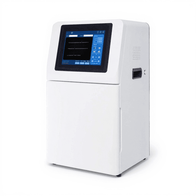
Chemiluminescence Imaging System
High-sensitivity chemiluminescence imaging system with 9-megapixel cooled camera for quantitative protein gel analysis and Western blot documentation.

Louise Corscadden, PhD
Director of Science · ConductScience
Ask Louise about Chemiluminescence Imaging System fit, setup, configuration, or quote prep.
Already working with us? Sign in to connect this with My Scientist.
Key Specifications
Full details →- Model fit
- Configured during quote
- SKU family
- SB-SCG-W3000
- Sizing
- 47.5 x 45.0 x 35.0 cm
- Ordering
- Online checkout and quote request available
- Category
- Gel & Protein Analysis
- Build notes
- Confirm accessories, station layout, and support needs before purchase
The SB-SCG-W3000 Chemiluminescence Imaging System is a high-performance detection platform designed for quantitative analysis of protein gels, Western blots, and other chemiluminescent assays in molecular biology research. The system features a 9-megapixel cooled camera with 3000×3000 resolution and thermoelectric cooling to -40°C below ambient temperature, enabling detection of low-abundance proteins and weak chemiluminescent signals with minimal background noise.
The instrument operates on the principle of detecting photons emitted during enzyme-substrate reactions, particularly horseradish peroxidase (HRP) catalyzed luminol oxidation commonly used in Western blotting protocols. The cooled sensor architecture reduces thermal noise while maximizing signal-to-noise ratio (42.2-47 dB), making it suitable for quantitative band intensity analysis and sensitive protein detection workflows. The system supports multiple imaging modes including real-time imaging, time accumulation, and automatic exposure optimization for diverse experimental requirements.
How It Works
Chemiluminescence imaging relies on the detection of photons emitted during specific enzyme-substrate reactions. In the most common application, horseradish peroxidase (HRP) conjugated to secondary antibodies catalyzes the oxidation of luminol in the presence of hydrogen peroxide, producing an excited intermediate that emits light at approximately 428 nm as it returns to the ground state. The intensity of light emission is directly proportional to the amount of target protein present.
The SB-SCG-W3000 captures these photons using a thermoelectrically cooled CMOS sensor with 3.76 μm pixel pitch and 16-bit dynamic range. Cooling to -40°C below ambient temperature reduces dark current to 0.0003 electrons per second per pixel, minimizing thermal noise that would otherwise interfere with weak signal detection. The high full-well capacity (50.5 ke- in low conversion gain mode) prevents pixel saturation during long exposures, while low readout noise (1.24-3.22 electrons) ensures detection of subtle signal variations.
The optical system focuses emitted photons onto the sensor array, where each photon generates photoelectrons that are converted to digital signal through analog-to-digital conversion. The 16-bit grayscale output provides 65,536 intensity levels, enabling precise quantitative measurements across a wide dynamic range suitable for both strong and weak chemiluminescent signals.
Features & Benefits
Instrument Type
- Imaging Systems
Application Area
- Protein Analysis
Automation Level
- fully-automated
camera_pixels
- 9 million pixels
camera_type
- high-sensitivity cooled camera
detection_technology
- chemiluminescence
imaging_modes
- Real-time Imaging, Time Imaging, Time Accumulation, Automatic Exposure
model_number
- SB-SCG-W3000
Research Domain
- Cancer Research
- Cell Biology
- Developmental Biology
- Immunology
- Molecular Biology
- Neuroscience
- Pharmaceutical QC
Weight
- 54.83 kg
Dimensions
- L: 47.5 mm
- W: 45.0 mm
- H: 35.0 mm
| Feature | This Product | Typical Alternative | Advantage |
|---|---|---|---|
| Sensor Resolution | 9 megapixel (3000×3000) | Entry-level systems often provide 1-6 megapixel sensors | Higher resolution enables more precise band analysis and better molecular weight determination accuracy. |
| Dynamic Range | 16-bit (65,536 grayscale levels) | Basic systems may offer 8-12 bit conversion | Wider dynamic range prevents signal clipping and enables accurate quantification across varied protein expression levels. |
| Cooling Performance | -40°C below ambient temperature | Non-cooled or limited cooling systems | Reduced thermal noise significantly improves detection of weak chemiluminescent signals and low-abundance proteins. |
| Exposure Time Range | 0.1ms to 1 hour | Limited exposure ranges in basic models | Flexible exposure times accommodate both strong and extremely weak signals without requiring multiple systems. |
| Signal-to-Noise Ratio | 42.2-47 dB depending on gain mode | Lower SNR in non-cooled systems | Superior noise performance enables detection of subtle protein expression changes and weak bands. |
| Imaging Modes | Real-time, time imaging, time accumulation, automatic exposure | Basic systems often provide limited automation | Multiple imaging modes streamline workflow and optimize results for different experimental conditions. |
The SB-SCG-W3000 combines high-resolution imaging with advanced cooling technology and flexible exposure control for sensitive chemiluminescent detection. The 9-megapixel sensor and 16-bit dynamic range support precise quantitative analysis, while multiple imaging modes and automatic exposure features enhance experimental efficiency.
Practical Tips
Perform dark field calibration each time the system is powered on or when ambient temperature changes significantly.
Why: Temperature variations affect dark current levels and baseline noise, impacting quantitative accuracy.
Clean the camera lens and sample chamber glass surfaces weekly with appropriate optical cleaning materials.
Why: Dust and residue on optical surfaces reduce light transmission and can create artifacts in images.
Use 2×2 or 3×3 binning for weak signals and 1×1 binning when maximum spatial resolution is required.
Why: Binning increases effective pixel size and sensitivity but reduces spatial resolution for band separation.
Verify signal linearity by capturing images at multiple exposure times for quantitative applications.
Why: Ensuring linear response across the dynamic range is critical for accurate protein quantification.
If images appear noisy, verify the cooling system is functioning and allow additional time for thermal equilibration.
Why: Inadequate cooling results in increased thermal noise that degrades image quality and sensitivity.
Optimize exposure time to utilize 50-80% of the dynamic range without pixel saturation.
Why: This range provides optimal signal-to-noise ratio while maintaining quantitative accuracy and avoiding clipping.
Ensure proper ventilation around the cooling system and avoid blocking air vents during operation.
Why: Adequate airflow is essential for thermoelectric cooling efficiency and prevents system overheating.
Document exposure settings and gain modes used for each experiment to ensure reproducible quantitative results.
Why: Consistent imaging parameters are essential for comparing results across different experiments and time points.
Setup Guide
What’s in the Box
- SB-SCG-W3000 Chemiluminescence Imaging System main unit
- Power adapter and cord
- USB communication cable
- Imaging software and drivers (CD/download)
- User manual and quick start guide
- Sample positioning guides (typical)
- Calibration certificate (typical)
Warranty
ConductScience provides a standard one-year manufacturer warranty covering parts and labor, with comprehensive technical support for installation, calibration, and troubleshooting assistance.
Compliance
What is the minimum detectable chemiluminescent signal with this system?
The system's sensitivity depends on exposure time and binning mode. With -40°C cooling and low readout noise (1.24 electrons in HCG mode), weak signals can be detected with extended exposure times up to 1 hour. Consult product datasheet for specific detection limits with different substrates.
How does the dual conversion gain mode affect quantitative analysis?
High conversion gain (HCG) mode provides lower readout noise (1.24e-) for sensitive detection, while low conversion gain (LCG) mode offers higher full-well capacity (50.5ke-) to prevent saturation. Choose based on expected signal strength and dynamic range requirements.
What gel formats and sizes are compatible with the imaging area?
The 11.28 mm × 11.28 mm target size accommodates mini-gels and membrane sections. For larger formats, consult product specifications for field of view limitations and positioning requirements.
How long does the camera require to reach operating temperature?
The thermoelectric cooling system typically requires 15-20 minutes to reach -40°C below ambient temperature. Full thermal equilibration may take longer in warm laboratory environments.
Can the system be used for fluorescence imaging applications?
This is a monochrome system optimized for chemiluminescence detection. For fluorescence applications, verify spectral sensitivity and excitation light compatibility with your specific fluorophores.
What data formats are supported for image export and analysis?
The system provides 16-bit grayscale images. Consult the software documentation for specific file formats and compatibility with third-party analysis software packages.
How does binning mode selection affect image quality and sensitivity?
1×1 binning provides maximum spatial resolution for detailed band analysis. 2×2 and 3×3 binning increase sensitivity at the cost of spatial resolution, useful for weak signal detection.
What maintenance procedures are required for optimal performance?
Regular dark field calibration, lens cleaning, and temperature monitoring are recommended. Consult the user manual for specific maintenance schedules and procedures to maintain detection sensitivity.
Have a question about this product?
Have a question? Just ask.
Send it over and we'll email you a personalized answer — no call, no scheduling.
Prefer to talk it through?
Accessories
Enhance your setup with compatible accessories