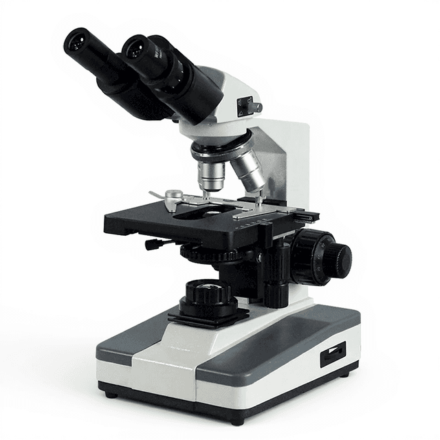
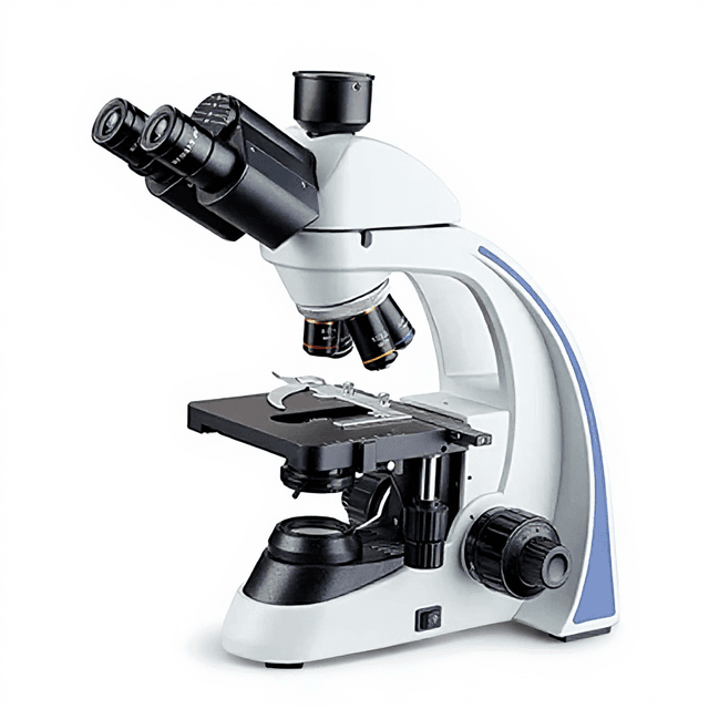
Fluorescence Biological Microscope BM-13CYII (Customized)
Research-grade fluorescence microscope with infinite optical system, apochromatic objectives 4X-100X, and magnification range 40X-1000X for cellular and molecular imaging applications.
| Automation Level | manual |
| Type | Magnification |
| Wide Plan Field Eyepiece | 10X |
| NA | Working distance |
| Infinite Apochromatic Fluoresecent Objective | 4X |
| 0.1 | 37.5mm |
The Fluorescence Biological Microscope BM-13CYII is a research-grade fluorescence microscope designed for advanced cellular and molecular imaging applications. Built on an infinite optical system, this microscope features apochromatic fluorescence objectives ranging from 4X to 100X with numerical apertures up to 1.25, providing high-resolution imaging capabilities across magnifications from 40X to 1000X. The system incorporates a four-position nosepiece for rapid objective changes and an Abbe condenser with iris diaphragm for optimal specimen illumination.
Equipped with wide-field 10X eyepieces and a precision mechanical stage with 75x50mm movement range, the microscope enables detailed examination of fluorescently-labeled specimens. The coaxial focus mechanism offers fine adjustment resolution to 0.002mm across a 30mm focusing range, supporting high-precision imaging requirements in research applications.
How It Works
Fluorescence microscopy operates on the principle of fluorescence excitation and emission. When fluorophores within the specimen are illuminated with light of a specific wavelength, they absorb this excitation energy and re-emit light at a longer wavelength. The microscope's optical system collects this emitted fluorescent light while filtering out the excitation wavelength, creating high-contrast images of labeled structures.
The infinite optical system eliminates spherical aberration by using parallel light rays between the objective and tube lens. This design allows for the insertion of additional optical components without affecting image quality. The apochromatic objectives correct for chromatic aberration across multiple wavelengths, ensuring sharp focus and accurate color reproduction essential for multi-fluorophore imaging.
The Abbe condenser with iris diaphragm provides uniform specimen illumination by focusing the light source onto the specimen plane. Proper condenser adjustment ensures optimal contrast and resolution by matching the numerical aperture of the condenser to the objective in use.
Features & Benefits
Automation Level
- manual
Type
- Magnification
Wide Plan Field Eyepiece
- 10X
NA
- Working distance
Infinite Apochromatic Fluoresecent Objective
- 4X
0.1
- 37.5mm
10X
- 0.25
40X
- 0.65
100X
- 1.25
Mechanical Tube Length
- Infinite Distance system
Total Magnification
- 40X~1000X
Nosepiece
- Fourtuple Nosepiece
Stage:
- Stage Size 160x140mm Moving Range 75x50mm
Focusing Range:
- 30mmCoaxial coarse and fine focus adjustment,Fine Focusing Scale0.002mm
Condenser
- Abbe N.A.1.25 Condenser With lris Diaphragm
Brand
- ConductScience
Research Domain
- Cancer Research
- Cell Biology
- Developmental Biology
- Histopathology
- Microbiology
- Neuroscience
Weight
- 29.98 kg
Dimensions
- L: 42.0 mm
- W: 43.6 mm
- H: 38.0 mm
Comparison Guide
| Feature | This Product | Typical Alternative | Advantage |
|---|---|---|---|
| Optical System Design | Infinite distance system | Entry-level models often use finite tube length systems | Allows insertion of additional optical components without affecting image quality and reduces spherical aberration. |
| Objective Correction | Apochromatic fluorescence objectives | Basic models typically offer achromatic objectives | Provides superior chromatic aberration correction essential for accurate multi-fluorophore imaging applications. |
| Magnification Range | 40X to 1000X total magnification | Limited magnification options in basic systems | Covers complete range from specimen overview to high-resolution detail examination in a single instrument. |
| Focus Precision | Fine focusing scale 0.002mm | Standard models offer less precise focus control | Enables precise specimen positioning critical for high-resolution fluorescence imaging and measurement accuracy. |
| Stage Movement | 75x50mm movement range on 160x140mm stage | Smaller stages with limited movement range | Accommodates larger specimens and provides extensive field mapping capabilities for comprehensive sample analysis. |
| Condenser Specifications | Abbe N.A. 1.25 condenser with iris diaphragm | Lower numerical aperture condensers in basic models | Matches the numerical aperture of high-magnification objectives for optimal resolution and contrast performance. |
This fluorescence microscope combines apochromatic objectives with an infinite optical system and precision mechanical components to deliver research-grade imaging capabilities. The system provides comprehensive magnification coverage with high numerical aperture objectives and precise focus control suitable for advanced cellular and molecular imaging applications.
Practical Tips
Verify objective parfocality by checking that specimens remain in focus when switching between objectives.
Why: Proper parfocality ensures efficient workflow and prevents specimen damage during magnification changes.
Clean objectives immediately after use, especially oil immersion lenses, using appropriate lens cleaning solutions and techniques.
Why: Residual immersion oil can degrade image quality and potentially damage objective lens coatings over time.
Match condenser numerical aperture to the objective in use by adjusting the iris diaphragm appropriately.
Why: Optimal condenser adjustment maximizes resolution and contrast while preventing excessive specimen heating.
If image quality degrades, check for contamination on eyepieces, objectives, and condenser surfaces before adjusting optical components.
Why: Dust and contamination are common causes of reduced image quality that can be easily resolved with proper cleaning.
Allow sufficient time for thermal equilibration when switching from low to high magnification objectives.
Why: Temperature changes can cause focus drift that affects measurement accuracy and image stability.
Use appropriate eye protection when working with fluorescence excitation sources and avoid prolonged direct exposure.
Why: Fluorescence excitation wavelengths can cause eye damage and specimen photobleaching with excessive exposure.
Store objectives in protective cases when not in use and maintain dust covers over the microscope.
Why: Proper storage prevents optical surface contamination and extends component lifetime.
Perform periodic alignment checks of the condenser and ensure proper centering with the optical axis.
Why: Misaligned condensers result in uneven illumination and reduced image quality across the field of view.
Setup Guide
What’s in the Box
- Fluorescence biological microscope main unit
- 10X wide-field eyepieces (pair)
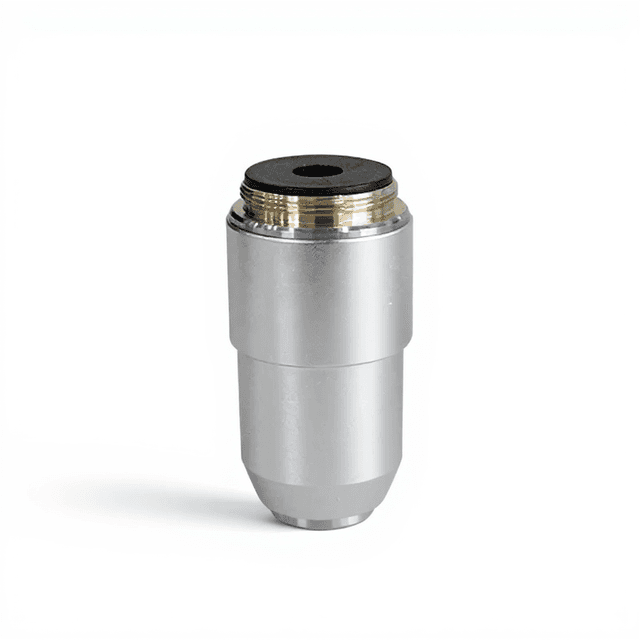
- Apochromatic fluorescence objectives (4X, 10X, 40X, 100X)
- Four-position nosepiece
- Abbe condenser with iris diaphragm
- Mechanical stage with specimen clips
- Power cord
- Dust cover
- User manual and documentation (typical)
- Objective lens cleaning kit (typical)
Warranty
ConductScience provides a standard one-year manufacturer warranty covering defects in materials and workmanship, with technical support for setup, operation, and maintenance guidance.
Compliance
What is the resolution limit at the highest magnification?
With the 100X oil immersion objective (N.A. 1.25), theoretical resolution approaches 0.2 micrometers, though practical resolution depends on specimen preparation and illumination conditions.
Can this microscope accommodate different fluorescence filter sets?
The infinite optical system design allows for insertion of fluorescence filter cubes, though specific filter compatibility should be verified with the technical specifications.
What type of specimens require the oil immersion objective?
The 100X oil immersion objective is essential for high-resolution examination of cellular ultrastructure, bacterial morphology, and detailed subcellular fluorescence localization studies.
How precise is the stage positioning for repeat measurements?
The mechanical stage provides reproducible positioning across the 75x50mm movement range, suitable for multi-field imaging and coordinate-based specimen mapping.
What maintenance is required for the optical components?
Regular cleaning of objectives and eyepieces with appropriate lens cleaning solutions, periodic condenser alignment verification, and protective storage when not in use.
Can this microscope be used for live cell imaging?
While the optical system supports live cell observation, extended imaging requires consideration of specimen heating and photobleaching under fluorescence excitation.
What is the working distance for routine screening?
The 4X objective offers 37.5mm working distance for initial specimen screening, while the 10X objective provides moderate magnification with sufficient working distance for most routine applications.
Have a question about this product?
Accessories
Enhance your setup with compatible accessories


Replacement Parts & Consumables


Frequently Bought Together
Upgrade Options
Related Products