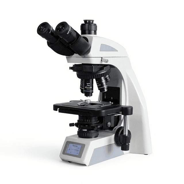
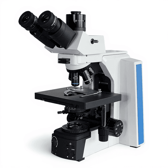
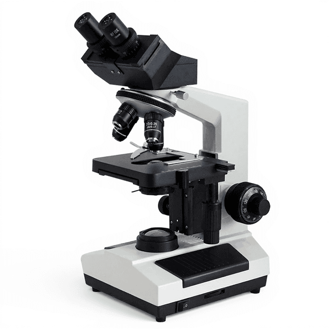
Infinity Trinocular Biological Microscope 40X~1000X with LED illumination
Infinity-corrected trinocular biological microscope with 40X-1000X magnification, LED illumination, and precision focusing for cellular and tissue analysis.

Louise Corscadden, PhD
Director of Science · ConductScience
Ask Louise about Infinity Trinocular Biological Microscope 40X~1000X with LED illumination fit, setup, configuration, or quote prep.
Already working with us? Sign in to connect this with My Scientist.
Key Specifications
Full details →- Model fit
- Configured during quote
- SKU family
- MCX-0002
- Sizing
- 4.9 x 6.6 x 6.6 cm
- Ordering
- Online checkout and quote request available
- Category
- Microscopy
- Build notes
- Confirm accessories, station layout, and support needs before purchase
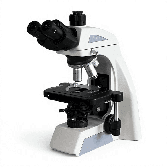
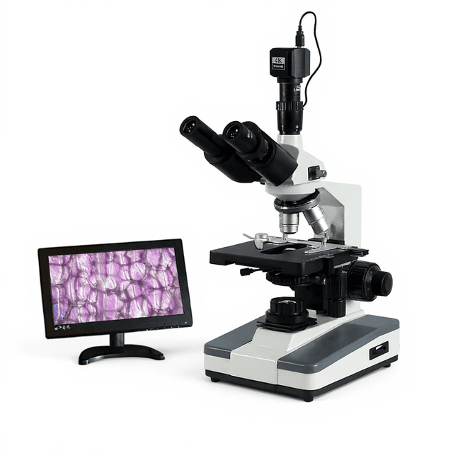
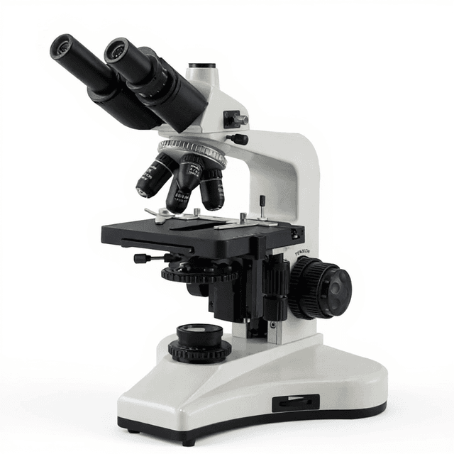
The Infinity Trinocular Biological Microscope delivers comprehensive optical performance for routine biological and medical research applications. This compound microscope features an infinity-corrected achromatic optical system with 40X to 1000X magnification range, providing researchers with the resolution and contrast needed for cellular and tissue examination. The trinocular head design enables simultaneous visual observation and image capture through the dedicated camera port.
Built with precision mechanics including coaxial coarse/fine focusing with 2 μm minimum division and a double-layer mechanical stage, this microscope supports quantitative measurements and extended observation sessions. The LED illumination system with brightness adjustment and color filters (green, yellow, blue) allows optimization for various staining techniques and contrast enhancement methods commonly used in biological microscopy.
How It Works
The microscope employs an infinity-corrected optical system where light from the specimen is collimated by the objective lens into parallel rays, then focused by a tube lens to form the image. This configuration eliminates spherical aberration and allows for the insertion of optical components (filters, beam splitters) in the parallel light path without affecting image quality. The plan achromatic objectives correct for field curvature and spherical aberration across the entire field of view.
Köhler illumination is achieved through the ABBE condenser (N.A. 1.25) with integrated diaphragm, ensuring even illumination across the specimen plane. The LED light source provides stable, cool illumination with adjustable intensity, while the color filters modify the spectral characteristics to enhance contrast for specific staining methods. The trinocular head splits the light path with an 80:20 ratio, directing 80% to the eyepieces for visual observation and 20% to the camera port for documentation.
Features & Benefits
Automation Level
- manual
Product Type
- Microscope
Microscope Type
- Biological / Compound
Viewing Head
- Trinocular
Application
- Clinical Research
- Life Science
Skill Level
- Intermediate
Brand
- ConductScience
Research Domain
- Cancer Research
- Cell Biology
- Clinical Diagnostics
- Developmental Biology
- Histopathology
- Microbiology
Weight
- 1.9 kg
Dimensions
- L: 4.9 mm
- W: 6.6 mm
- H: 6.6 mm
| Feature | This Product | Typical Alternative | Advantage |
|---|---|---|---|
| Optical System Design | Infinity-corrected achromatic system | Entry-level models often use finite tube length systems with standard achromat objectives | Infinity correction allows addition of optical components without changing magnification and reduces optical aberrations for more accurate measurements. |
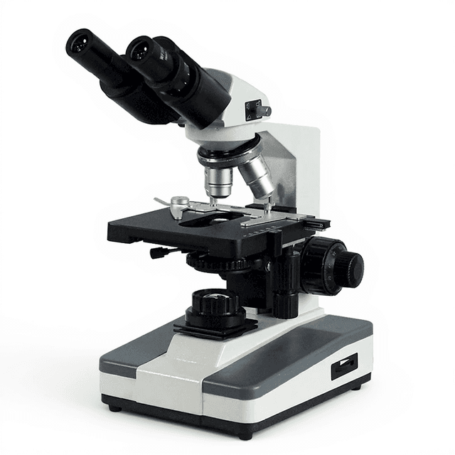
| Head Configuration | Trinocular head with 80:20 beam splitting | Basic models typically offer binocular heads only | Simultaneous observation and documentation capability essential for research protocols requiring image capture and analysis. |
| Objective Quality | Plan achromatic objectives providing flat field imaging | Standard achromat objectives with field curvature limitations | Plan correction ensures sharp focus across the entire field of view, critical for quantitative measurements and photomicrography. |
| Focusing Precision | Coaxial focusing with 2 μm minimum division | Basic systems often provide 10-20 μm focusing increments | Fine focusing precision enables critical focus applications and reduces drift during extended observation periods. |
| Condenser Specification | ABBE condenser with N.A. 1.25 | Lower-end models may have N.A. 0.9-1.0 condensers | Higher numerical aperture supports full resolution potential of 100X oil immersion objectives for maximum detail resolution. |
| Illumination System | LED with adjustable brightness and color filters | Tungsten halogen lamps with limited intensity control | LED provides stable illumination with extended lamp life and consistent color temperature for reproducible imaging protocols. |
This infinity-corrected microscope provides research-grade optical performance with trinocular documentation capability, plan achromatic objectives, and precision focusing typically found in higher-end systems. The LED illumination and comprehensive filter set support diverse staining protocols and imaging requirements for biological research applications.
Practical Tips
Perform Köhler illumination setup each time you change objectives or specimens to maintain optimal resolution and contrast.
Why: Proper illumination alignment is critical for achieving the theoretical resolution limit of each objective.
Clean objective lenses with lens tissue and appropriate solvents immediately after oil immersion use to prevent oil hardening.
Why: Dried immersion oil can permanently damage objective coatings and reduce image quality.
Always start examination with the lowest magnification objective and progress systematically to higher magnifications.
Why: This approach protects objectives from damage and allows efficient specimen survey before detailed examination.
If image appears dim or uneven, check condenser centering and aperture diaphragm position before adjusting illumination intensity.
Why: Optical misalignment causes illumination problems that cannot be corrected by simply increasing lamp brightness.
Use the color filters to enhance contrast for specific staining methods rather than relying solely on digital image processing.
Why: Optical contrast enhancement preserves more image information than post-acquisition digital manipulation.
Allow LED illumination to stabilize for 5-10 minutes before critical measurements to ensure consistent color temperature and intensity.
Why: Temperature stabilization prevents measurement drift and ensures reproducible imaging conditions.
Use the mechanical stage locks when switching between observation and documentation to maintain specimen position.
Why: Stage movement during mode switching can result in loss of the specific field being examined or documented.
Calibrate the eyepiece reticle or camera system using a stage micrometer for each objective used in quantitative measurements.
Why: Each objective has a different magnification factor that affects measurement accuracy and requires individual calibration.
Setup Guide
What’s in the Box
- Infinity trinocular biological microscope
- WF10X/18mm eyepieces (pair)
- Plan achromatic objectives: 4X, 10X, 40X, 100X (oil)
- External power adapter (100-240V AC to 12V DC)
- Color filter set (green, yellow, blue)
- Immersion oil for 100X objective (typical)
- Dust cover (typical)
- User manual and documentation (typical)
- Power cord appropriate for region (typical)
Warranty
ConductScience provides a standard one-year manufacturer warranty covering defects in materials and workmanship, with technical support for setup and operation.
Compliance
References
Background reading relevant to this product:
What is the numerical aperture limit for high-resolution imaging with this system?
The ABBE condenser provides N.A. 1.25, which matches the requirements for the 100X oil immersion objective and supports resolution down to approximately 0.22 micrometers with proper Köhler illumination setup.
Can this microscope accommodate immunofluorescence applications?
The system includes excitation filters and trinocular design suitable for fluorescence documentation, though specific filter cubes for immunofluorescence wavelengths would need to be added separately based on fluorophore requirements.
What camera mounting options are available for the trinocular port?
The trinocular port uses standard C-mount threading and receives 20% of the light path. Camera selection depends on sensor size and resolution requirements; consult product datasheet for specific mounting specifications.
How does the infinity correction benefit quantitative measurements?
Infinity correction eliminates magnification changes when adding filters or beam splitters in the optical path, ensuring consistent measurements across different observation modes and maintaining calibration accuracy.
What maintenance schedule is recommended for the LED illumination system?
LED systems typically require minimal maintenance with 20,000+ hour lifespans. Regular cleaning of illumination optics and periodic intensity calibration are recommended for quantitative applications.
Can the mechanical stage accommodate standard slide formats and larger specimens?
The 155×142mm stage accommodates standard 75×25mm microscope slides as well as larger specimens like Petri dishes, with 76×50mm X-Y movement range for systematic examination.
What focusing precision is achievable for critical focus applications?
The coaxial fine focus provides 2 μm minimum division with tension adjustment and upper limit stop, suitable for precise focus positioning in high-magnification applications and Z-stack imaging.
How does this compare to phase contrast microscopy systems?
This brightfield system excels for stained specimens and routine histology, while phase contrast systems better visualize unstained living cells. Phase contrast capabilities would require additional condenser and objective modifications.
Have a question about this product?
Have a question? Just ask.
Send it over and we'll email you a personalized answer — no call, no scheduling.
Prefer to talk it through?
Accessories
Enhance your setup with compatible accessories