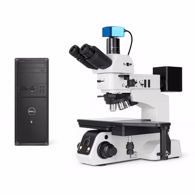
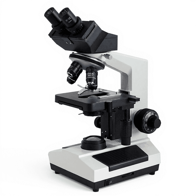
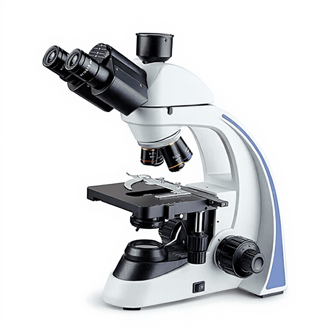
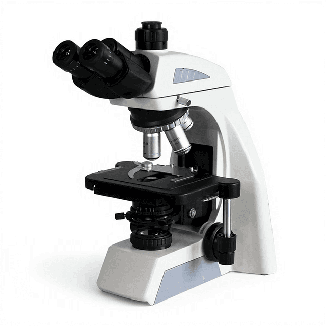
Binocular/Trinocular Microscope Laboratory Clinical Biology Biological Microscope for Lab Live Blood Analysis Use
Brightfield compound microscope with 40X-1600X magnification range, featuring binocular/trinocular head, Abbe condenser, and halogen illumination for clinical biology and live blood analysis applications.

Louise Corscadden, PhD
Director of Science · ConductScience
Ask Louise about Binocular/Trinocular Microscope Laboratory Clinical Biology Biological Microscope for Lab Live Blood Analysis Use fit, setup, configuration, or quote prep.
Already working with us? Sign in to connect this with My Scientist.
Key Specifications
Full details →- Model fit
- Configured during quote
- SKU family
- MCX-0004
- Sizing
- 4.9 x 6.6 x 6.6 cm
- Ordering
- Online checkout and quote request available
- Category
- Microscopy
- Build notes
- Confirm accessories, station layout, and support needs before purchase
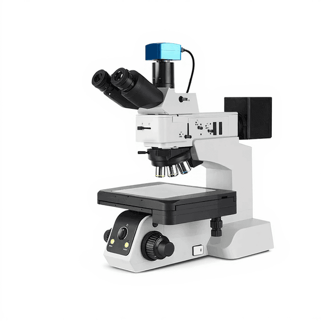
The ConductScience Binocular/Trinocular Microscope represents a versatile brightfield compound microscope designed for clinical biology and laboratory applications. This instrument provides magnifications from 40X to 1600X through its combination of WF10X (Ø18mm) and WF16X (Ø11mm) eyepieces with a quadruple nosepiece system. The microscope features a 360° rotatable binocular/trinocular head inclined at 30° with adjustable interpupillary distance (48-75mm) for ergonomic viewing.
The optical system incorporates an Abbe condenser (N.A. 1.25) with rack and pinion adjustment, coaxial coarse and fine focusing with 2μm minimum fine focus division, and a double-layer mechanical stage (135mm x 140mm) with 70mm x 50mm movement range. Illumination is provided by a 6V 20W halogen lamp with brightness control, supported by blue, yellow, and green filters for enhanced contrast in various specimen types.
How It Works
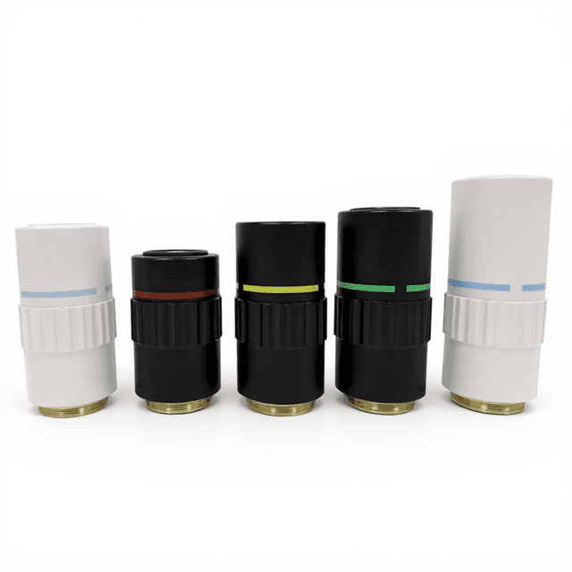
This compound microscope operates on the principle of brightfield illumination, where light from the halogen source passes through the specimen and into the objective lens system. The Abbe condenser (N.A. 1.25) focuses illumination onto the specimen plane, while the iris diaphragm controls light intensity and contrast. The quadruple nosepiece allows rapid switching between different magnification objectives, though only the 4X objective specifications (N.A. 0.10, working distance 37.50mm) are provided in the current configuration.
The coaxial focusing system provides both coarse and fine adjustments, with the fine focus mechanism offering 2μm precision over a 30mm range. The double-layer mechanical stage enables precise specimen positioning through X-Y movement controls spanning 70mm x 50mm. Light filtering through blue, yellow, and green filters enhances contrast for different specimen types and staining protocols.
The binocular/trinocular head configuration allows for comfortable viewing through the eyepieces while maintaining the option for camera attachment through the trinocular port. The 360° rotation capability accommodates multiple users and various working positions.
Features & Benefits
Automation Level
- manual
Product Type
- Microscope
Microscope Type
- General Purpose
Viewing Head
- Trinocular
Application
- Clinical Research
- Laboratory Research
- Life Science
Skill Level
- Intermediate
Brand
- ConductScience
Research Domain
- Cell Biology
- Clinical Diagnostics
- Histopathology
- Microbiology
- Pharmaceutical QC
Weight
- 1.9 kg
Dimensions
- L: 4.9 mm
- W: 6.6 mm
- H: 6.6 mm
| Feature | This Product | Typical Alternative | Advantage |
|---|---|---|---|
| Condenser Numerical Aperture | N.A. 1.25 Abbe condenser with diaphragm | Entry-level models often provide N.A. 1.25 or lower | Higher numerical aperture improves resolution and light-gathering capability for detailed specimen analysis. |
| Focusing Precision | 2μm minimum fine focus division | Basic models may offer 5-10μm precision | Finer focus control enables precise imaging at high magnifications where depth of field is minimal. |
| Head Configuration | 360° rotatable binocular/trinocular head | Fixed-angle heads are common in basic models | Rotation capability accommodates multiple users and various working positions in shared laboratory settings. |
| Stage Design | Double-layer mechanical stage (70mm x 50mm range) | Single-layer or fixed stages with limited movement | Enhanced specimen positioning control enables systematic scanning and precise region-of-interest location. |
| Illumination Control | Brightness control with 3-filter set | Fixed intensity or limited filter options | Adjustable illumination and contrast filtering optimize visualization for different specimen types and staining protocols. |
| Power Compatibility | Wide voltage range (AC 85-265V, 50-60Hz) | Fixed voltage requirements vary by region | Universal power compatibility simplifies installation in international laboratory environments. |
This microscope offers research-grade optical performance with Abbe condenser, precision focusing, and versatile head configuration. The combination of mechanical stage control, illumination flexibility, and wide power compatibility provides reliable performance for clinical and research applications.
Practical Tips
Verify interpupillary distance setting matches your eye spacing and adjust eyepiece diopters for balanced focus between both eyes.
Why: Proper calibration reduces eye strain and ensures accurate specimen observation during extended use.
Clean optical surfaces with appropriate lens paper and solution, avoiding household cleaners that can damage coatings.
Why: Regular optical maintenance preserves image quality and prevents permanent damage to expensive optical elements.
Start observation at low magnification (4X objective) and progressively increase magnification after locating the region of interest.
Why: This systematic approach prevents specimen loss and reduces time spent searching at high magnifications with narrow fields of view.
If focus seems soft at high magnifications, check that the condenser height is properly adjusted and the iris diaphragm is not completely closed.
Allow the halogen lamp to warm up for several minutes before critical observations to ensure stable illumination intensity.
Why: Lamp stabilization prevents brightness variations that can affect image quality and photographic documentation.
Never force mechanical adjustments and ensure specimen slides are properly secured in stage clips before movement.
Why: Gentle handling prevents damage to precision mechanisms and avoids specimen contamination or breakage.
Use appropriate color filters (blue for enhanced nuclear detail, yellow for general contrast) based on specimen type and staining.
Why: Filter selection optimizes contrast for specific cellular structures and improves diagnostic accuracy.
Store the microscope with dust cover and verify all mechanical movements operate smoothly during routine checks.
Why: Environmental protection and regular mechanical verification prevent contamination and ensure consistent performance.
Setup Guide
What’s in the Box
- Binocular/trinocular microscope body
- WF10X eyepiece (Ø18mm) (typical)
- WF16X eyepiece (Ø11mm) (typical)
- 4X achromatic objective (N.A. 0.10)
- Blue, yellow, and green filters
- External power supply (AC 85-265V)
- User manual and optical cleaning cloth (typical)
- Dust cover (typical)
Warranty
ConductScience provides a standard one-year manufacturer warranty covering optical and mechanical components, with technical support for setup and operational guidance.
Compliance
References
Background reading relevant to this product:
What is the numerical aperture of the condenser and how does it affect resolution?
The Abbe condenser provides N.A. 1.25, which matches or exceeds most objective numerical apertures for optimal resolution. This ensures proper light cone angle for maximum resolving power across the magnification range.
Can this microscope accommodate both brightfield and phase contrast techniques?
The current configuration is optimized for brightfield microscopy with halogen illumination and color filters. Phase contrast capabilities would require additional phase objectives and condenser components not included in this specification.
What is the working distance of the 4X objective and why does this matter?
The 4X objective provides 37.50mm working distance, allowing examination of thick specimens and providing clearance for slide manipulation. Higher magnification objectives typically have shorter working distances.
How precise is the fine focusing mechanism for high-magnification work?
The fine focus mechanism offers 2μm minimum division across a 30mm range, providing the precision necessary for critical focusing at magnifications up to 1600X.
What specimen thickness can be accommodated with the mechanical stage?
The double-layer mechanical stage design accommodates standard microscope slides and specimens up to the limits of the 37.50mm working distance of the 4X objective. Consult specifications for higher magnification objectives.
Is the illumination system suitable for digital imaging applications?
The 6V 20W halogen lamp with brightness control provides stable illumination suitable for visual observation. For quantitative imaging, consider color temperature specifications and potential need for additional stabilization.
How does the trinocular port affect the light path for visual observation?
The trinocular configuration typically uses a beam splitter that directs a portion of light to the camera port while maintaining full illumination to the binocular eyepieces for simultaneous viewing and imaging.
What maintenance is required for the mechanical stage and focusing mechanisms?
Regular cleaning of stage surfaces and periodic lubrication of focusing mechanisms according to manufacturer specifications ensures smooth operation and prevents specimen contamination.
Have a question about this product?
Have a question? Just ask.
Send it over and we'll email you a personalized answer — no call, no scheduling.
Prefer to talk it through?
Accessories
Enhance your setup with compatible accessories

Frequently Bought Together
Upgrade Options
Related Products