Pryer Endoscopic Objective 30x
Miniaturized 30x endoscopic objective with 0.8 μm resolution and 8.35 mm imaging depth for high-resolution deep tissue microscopy through a 3.0 mm diameter probe.

Louise Corscadden, PhD
Director of Science · ConductScience
Ask Louise about Pryer Endoscopic Objective 30x fit, setup, configuration, or quote prep.
Already working with us? Sign in to connect this with My Scientist.
Key Specifications
Full details →- Model fit
- Mouse, Rat, Zebrafish
- SKU family
- MCI-EDO-30X
- Sizing
- 0.12 x 25.0 x 45.0 cm
- Ordering
- Online checkout and quote request available
- Category
- Microscopy
- Build notes
- Confirm accessories, station layout, and support needs before purchase
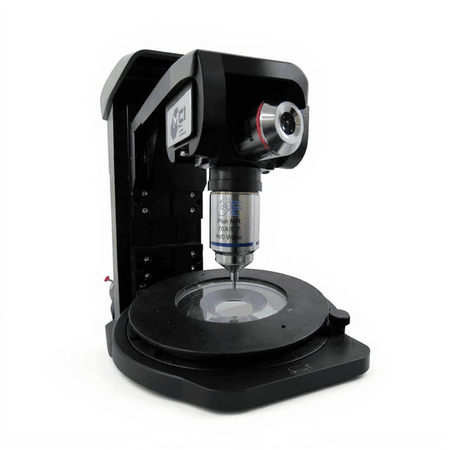
The Pryer Endoscopic Objective 30x is a miniaturized gradient index (GRIN) lens system designed for high-resolution endoscopic imaging applications. This objective provides 0.8 μm resolution with a 0.5 numerical aperture, enabling cellular-level visualization through a compact 3.0 mm diameter probe. The system operates across a 400-1080 nm spectral range, supporting both fluorescence and brightfield imaging modalities.
With an 8.35 mm total imaging depth and 500 μm field of view, this endoscopic objective enables deep tissue microscopy in confined spaces where conventional objectives cannot reach. The miniaturized form factor makes it particularly suitable for in vivo imaging applications, microendoscopy studies, and situations requiring optical access through small apertures or working channels.
How It Works
The Pryer Endoscopic Objective 30x utilizes gradient index (GRIN) lens technology to achieve high numerical aperture imaging through a miniaturized form factor. GRIN lenses contain a radially varying refractive index profile that focuses light without requiring curved surfaces, enabling the fabrication of rod-shaped objectives with diameters as small as 3.0 mm.
Light collected by the objective is focused through the GRIN lens system, which maintains a 0.5 numerical aperture while providing 0.8 μm resolution across the 400-1080 nm working band. The 7.85 mm lens length combined with a 0.5 mm working distance provides an effective imaging depth of 8.35 mm, allowing visualization deep within tissue samples or confined experimental chambers.
The 500 μm field of view enables simultaneous observation of multiple cellular structures while maintaining the resolution necessary for subcellular feature discrimination. This combination of high resolution, extended working distance, and compact geometry makes the system particularly effective for applications requiring optical access through small apertures or in spatially constrained environments.
Features & Benefits
imaging-depth
- 7.85 mm + 0.5 mm = 8.35 mm
numerical-aperture
- 0.5
tip-dimensions
- 7.85 mm length / 3.0 mm diameter
working-band
- 400 - 1080 nm
field-of-view
- 500 um
Automation Level
- manual
Accuracy
- 0.8 um
Research Domain
- Cancer Research
- Cardiovascular
- Cell Biology
- Developmental Biology
- Histopathology
- Neuroscience
Species
- Mouse
- Rat
- Zebrafish
Weight
- 22.05 kg
Dimensions
- L: 0.12 mm
- W: 25.0 mm
- H: 45.0 mm
| Feature | This Product | Typical Alternative | Advantage |
|---|---|---|---|
| Numerical Aperture | 0.5 NA | Entry-level endoscopic objectives often provide lower numerical apertures | Higher light collection and resolution capability enables better image quality in challenging deep tissue applications |
| Resolution | 0.8 μm resolution | Basic endoscopic systems may offer 1-2 μm resolution | Subcellular feature discrimination allows detailed analysis of cellular morphology and fine structures |
| Imaging Depth | 8.35 mm total imaging depth | Standard objectives typically limited to shorter working distances | Extended depth enables imaging of structures beyond the reach of conventional microscopy systems |
| Spectral Range | 400-1080 nm working band | Some systems limited to visible wavelengths only | Broad spectral coverage supports both fluorescence and near-infrared imaging applications |
| Probe Diameter | 3.0 mm diameter | Larger diameter probes may restrict access to confined spaces | Compact geometry enables imaging through small apertures and working channels |
The Pryer Endoscopic Objective 30x combines 0.5 numerical aperture with 8.35 mm imaging depth in a 3.0 mm diameter probe, providing 0.8 μm resolution across a 400-1080 nm spectral range. This combination of high optical performance and compact geometry makes it well-suited for deep tissue imaging applications requiring cellular-level resolution.
Practical Tips
Regularly verify the 500 μm field of view calibration using stage micrometers to ensure accurate spatial measurements.
Why: Field of view accuracy is critical for quantitative analysis and proper interpretation of cellular spacing and morphology.
Store the objective with protective caps and avoid touching the lens tip to prevent contamination and optical degradation.
Why: The small diameter optical surfaces are particularly susceptible to contamination that can significantly impact image quality.
Maintain the 0.5 mm working distance consistently to ensure optimal resolution across the entire imaging session.
Why: Working distance variations directly affect focus quality and resolution performance in high numerical aperture systems.
If resolution appears degraded, check for contamination on the objective tip and verify proper mounting thread engagement.
Why: Small particles or improper mounting can significantly impact the high-resolution capabilities of miniaturized optical systems.
Use appropriate illumination intensity within the 400-1080 nm range to maximize signal-to-noise ratio without photobleaching.
Why: The high numerical aperture enables efficient light collection, allowing lower illumination levels that preserve sample viability.
When working with laser illumination sources, ensure proper safety protocols for the extended working distance configuration.
Why: The 8.35 mm imaging depth may require different laser safety considerations compared to conventional short working distance objectives.
Setup Guide
What’s in the Box
- Pryer Endoscopic Objective 30x
- Protective lens cap (typical)
- Technical specification sheet (typical)
- User manual (typical)
Warranty
ConductScience provides a 1-year manufacturer warranty covering defects in materials and workmanship, with technical support for optical performance and compatibility questions.
Compliance
What is the minimum working distance required for this objective?
The objective provides a 0.5 mm working distance, requiring this minimum spacing between the lens tip and sample surface for proper focus.
Can this objective be used with standard fluorescence filter sets?
Yes, the 400-1080 nm working band is compatible with most common fluorophores and standard filter cube configurations used in fluorescence microscopy.
What mounting thread specification does this objective use?
Consult the product datasheet for specific mounting thread dimensions and compatibility with your imaging system.
How does the 500 μm field of view compare to conventional objectives?
The field of view is smaller than standard objectives but optimized for cellular-level imaging through the miniaturized probe geometry required for endoscopic applications.
What is the maximum tissue penetration depth achievable?
The 8.35 mm imaging depth represents the optical working distance, while actual tissue penetration depends on sample transparency and scattering properties.
Is this objective compatible with two-photon imaging systems?
The 400-1080 nm working band includes near-infrared wavelengths commonly used in two-photon microscopy, though specific system compatibility should be verified.
How should the objective be cleaned and maintained?
Clean optical surfaces only with lens-safe solvents and lint-free materials, handling the objective by mounting threads to avoid damaging the precision optical elements.
Have a question about this product?
Have a question? Just ask.
Send it over and we'll email you a personalized answer — no call, no scheduling.
Prefer to talk it through?
Upgrade Options
Related Products