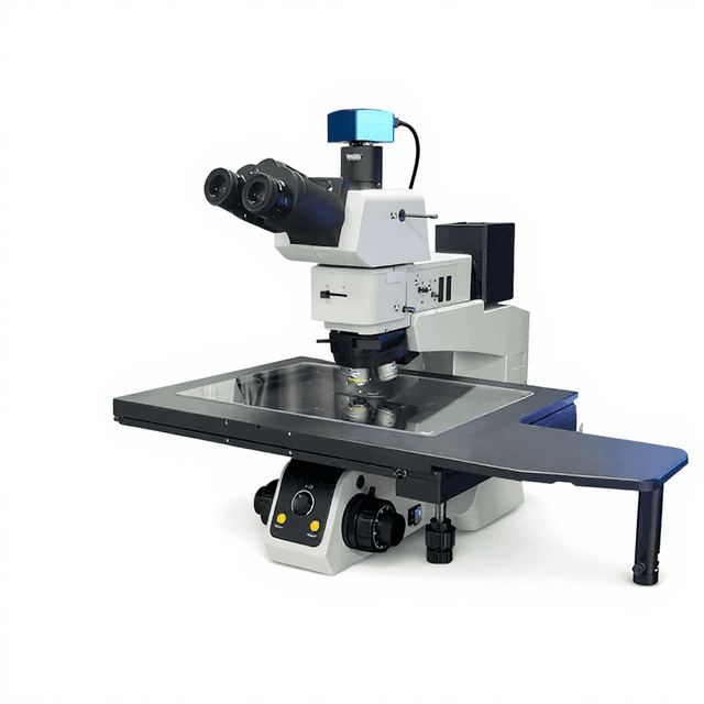
Pryer Endoscopic Objective 70x
High-resolution 70x endoscopic objective with 0.7 numerical aperture and 0.5 μm resolution for detailed microscopic imaging in confined spaces.

Louise Corscadden, PhD
Director of Science · ConductScience
Ask Louise about Pryer Endoscopic Objective 70x fit, setup, configuration, or quote prep.
Already working with us? Sign in to connect this with My Scientist.
Key Specifications
Full details →- Model fit
- Configured during quote
- SKU family
- MCI-EDO-70X
- Sizing
- 0.08 x 25.0 x 45.0 cm
- Ordering
- Online checkout and quote request available
- Category
- Microscopy
- Build notes
- Confirm accessories, station layout, and support needs before purchase
The Pryer Endoscopic Objective 70x is a high-resolution imaging component designed for endoscopic microscopy applications requiring detailed visualization at the cellular and subcellular level. With a numerical aperture of 0.7 and 0.5 μm resolution capability, this objective enables researchers to capture fine morphological details in live tissue preparations and biological specimens.
The objective features a compact 2.1 mm diameter tip with 5.87 mm working length, providing an imaging depth of up to 6.07 mm. The broad spectral response from 400-1080 nm supports both visible light and near-infrared imaging modalities, while the 200 μm field of view balances spatial coverage with high magnification detail capture.
How It Works
The Pryer Endoscopic Objective 70x operates on the principle of high-numerical-aperture light collection and focusing within a miniaturized optical system. The 0.7 numerical aperture design maximizes light-gathering capability while maintaining the compact form factor required for endoscopic applications. Light from the specimen is collected through the objective's front lens elements and focused through a series of precisely aligned optical components within the 2.1 mm diameter housing.
The objective's optical design incorporates correction elements to minimize spherical and chromatic aberrations across the 400-1080 nm working band. This broad spectral response enables multi-wavelength imaging techniques including fluorescence microscopy with various fluorophores and near-infrared imaging applications. The 5.87 mm working distance allows imaging through protective windows or within tissue while maintaining the 0.5 μm resolution capability.
Image formation occurs through the combination of the objective's magnification power and the numerical aperture's light-collecting efficiency. The 200 μm field of view represents the practical imaging area at the specimen plane, balancing magnification with spatial coverage for comprehensive sample examination.
Features & Benefits
imaging-depth
- 5.87 mm + 0.2 mm = 6.07 mm
numerical-aperture
- 0.7
tip-dimensions
- 5.87 mm length / 2.1 mm diameter
working-band
- 400 - 1080 nm
field-of-view
- 200 um
Accuracy
- 0.5 um
Research Domain
- Cell Biology
- Developmental Biology
- Histopathology
- Materials Science
- Microbiology
- Neuroscience
Weight
- 44.09 kg
Dimensions
- L: 0.08 mm
- W: 25.0 mm
- H: 45.0 mm
| Feature | This Product | Typical Alternative | Advantage |
|---|---|---|---|
| Numerical Aperture | 0.7 | Entry-level endoscopic objectives often offer 0.4-0.5 numerical aperture | Higher numerical aperture provides improved light collection and image brightness for better visualization in low-light conditions. |
| Resolution Capability | 0.5 μm | Basic endoscopic systems may achieve 1-2 μm resolution | Sub-micrometer resolution enables detailed cellular and subcellular structure visualization critical for biological research. |
| Spectral Range | 400-1080 nm | Standard objectives may be limited to visible spectrum only | Extended near-infrared capability supports advanced imaging techniques including multi-wavelength fluorescence applications. |
| Working Distance | 5.87 mm | High magnification objectives typically offer shorter working distances | Extended working distance allows imaging through protective windows and within tissue samples while maintaining high resolution. |
| Field of View | 200 μm | Varies by design and magnification | Optimized field size balances magnification with spatial coverage for comprehensive specimen examination. |
The Pryer Endoscopic Objective 70x combines high numerical aperture performance with the compact design required for confined space imaging. The broad spectral response and extended working distance provide operational flexibility while maintaining sub-micrometer resolution capability essential for detailed biological and materials research applications.
Practical Tips
Verify resolution performance using calibrated test targets or known cellular structures before critical imaging sessions.
Why: Ensures the objective is performing to specifications and helps identify any optical degradation.
Clean optical surfaces only with appropriate lens cleaning solutions and lint-free tissues to prevent scratching.
Why: Improper cleaning can permanently damage the optical coatings and degrade image quality.
Maintain consistent working distance of 5.87 mm for optimal image quality and resolution performance.
Why: Deviation from the designed working distance reduces resolution and can introduce optical aberrations.
If images appear dim or lack contrast, verify that illumination wavelength falls within the 400-1080 nm working band.
Why: Illumination outside the specified spectral range may not be efficiently transmitted through the objective optics.
Use appropriate exposure times to fully utilize the 0.7 numerical aperture light-gathering capability without oversaturation.
Why: Proper exposure maximizes signal-to-noise ratio while preventing pixel saturation that can obscure fine details.
Handle the objective by the body, never by the tip, and use protective caps when not in use.
Why: The precision optics and compact design make the objective susceptible to damage from mechanical stress or contamination.
Allow the objective to equilibrate to room temperature before use if stored in different environmental conditions.
Why: Temperature changes can affect optical alignment and image quality until thermal equilibrium is reached.
Store the objective in a clean, dry environment with protective caps installed on both ends.
Why: Proper storage prevents dust accumulation and moisture damage that can degrade optical performance.
Setup Guide
What’s in the Box
- Pryer Endoscopic Objective 70x
- Protective lens caps (typical)
- Optical cleaning cloth (typical)
- User manual and specifications sheet (typical)
Warranty
ConductScience provides a standard one-year manufacturer warranty covering defects in materials and workmanship, with technical support for installation and operational guidance.
Compliance
What is the minimum feature size this objective can resolve?
The objective provides 0.5 μm resolution, enabling visualization of cellular organelles and subcellular structures.
Can this objective be used for fluorescence imaging?
Yes, the 400-1080 nm working band supports common fluorophores including FITC, rhodamine, and near-infrared dyes.
What is the working distance limitation for specimen positioning?
The objective requires specimens to be positioned at 5.87 mm working distance, with a maximum imaging depth of 6.07 mm.
How does the 2.1 mm diameter compare to standard microscope objectives?
The compact diameter enables access through narrow channels and confined spaces not accessible to conventional larger objectives.
What field of view can I expect at this magnification?
The objective provides a 200 μm field of view, suitable for examining multiple cells or tissue regions simultaneously.
Is this objective compatible with standard microscope systems?
Compatibility depends on the mounting interface; consult product datasheet for specific threading and adapter requirements.
What maintenance is required for optimal performance?
Regular cleaning of optical surfaces and proper storage with protective caps will maintain image quality and prevent contamination.
Have a question about this product?
Have a question? Just ask.
Send it over and we'll email you a personalized answer — no call, no scheduling.
Prefer to talk it through?
Accessories
Enhance your setup with compatible accessories
Upgrade Options
Related Products