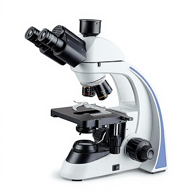
40X~1000X Laboratory Microscope Biological Laboratory Microscope for Research with Display
Professional compound microscope with 40X-1000X magnification range, trinocular head design, and precision coaxial focusing system for biological research applications.

Louise Corscadden, PhD
Director of Science · ConductScience
Ask Louise about 40X~1000X Laboratory Microscope Biological Laboratory Microscope for Research with Display fit, setup, configuration, or quote prep.
Already working with us? Sign in to connect this with My Scientist.
Key Specifications
Full details →- Model fit
- Configured during quote
- SKU family
- MCX-0008
- Sizing
- 4.9 x 6.6 x 6.6 cm
- Ordering
- Online checkout and quote request available
- Category
- Microscopy
- Build notes
- Confirm accessories, station layout, and support needs before purchase
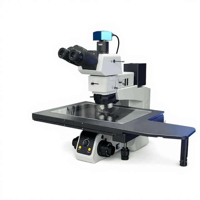
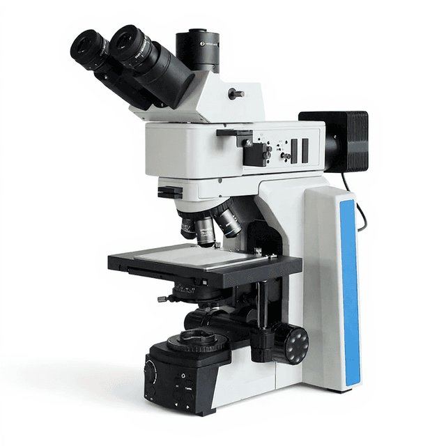
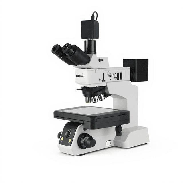
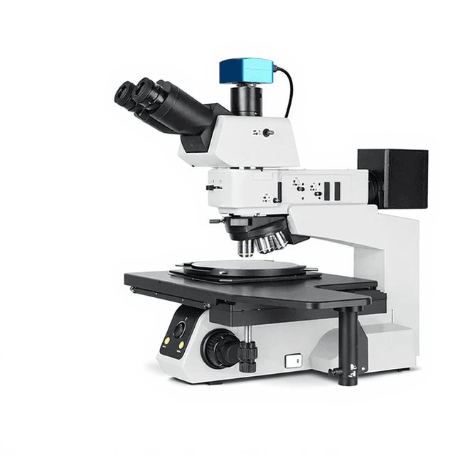
The MCX-0008 is a professional-grade compound microscope designed for biological research applications requiring magnifications from 40X to 1000X. This trinocular system features infinity-corrected plan achromatic objectives with numerical apertures optimized for high-resolution imaging across the visible spectrum. The instrument incorporates a quintuple nosepiece with precision ball bearing positioning, coaxial coarse/fine focusing with 2 μm minimum increments, and an ABBE condenser system with 1.25 numerical aperture for optimal specimen illumination.
The integrated 3W S-LED illumination system provides consistent, adjustable brightness with digital readout via LCD display. The double-layer mechanical stage offers 78mm x 54mm specimen positioning range with precision controls. The trinocular head design accommodates both binocular observation and simultaneous documentation through the third port, with 360° rotation and 30° inclined viewing angle for ergonomic operation during extended observation sessions.
How It Works
The microscope operates on transmitted light brightfield microscopy principles, where specimens are illuminated from below using the integrated LED system. Light passes through the ABBE condenser (N.A. 1.25), which focuses and concentrates the illumination onto the specimen plane. The condenser features rack and pinion adjustment with an aperture diaphragm for controlling light cone angle and contrast optimization.
The infinity-corrected optical system employs plan achromatic objectives that form parallel light rays, which are then focused by a tube lens to create the intermediate image. This design eliminates spherical and chromatic aberrations while maintaining consistent magnification and field flatness across the entire viewing area. The coaxial focusing mechanism provides both coarse (37.7mm range) and fine focusing (2 μm minimum increments) through precision gear reduction systems.
The trinocular head incorporates a beam splitter that directs light simultaneously to both eyepieces and the documentation port, enabling real-time observation while capturing images or video. The 360° rotatable head with 47-78mm interpupillary adjustment accommodates multiple users and viewing angles without compromising optical performance.
Features & Benefits
Automation Level
- manual
Product Type
- Microscope
Microscope Type
- Biological / Compound
Application
- Clinical Research
- Laboratory Research
- Life Science
Skill Level
- Intermediate
Brand
- ConductScience
Research Domain
- Cancer Research
- Cell Biology
- Clinical Diagnostics
- Developmental Biology
- Food Science
- Histopathology
- Materials Science
- Microbiology
Weight
- 1.9 kg
Dimensions
- L: 4.9 mm
- W: 6.6 mm
- H: 6.6 mm
| Feature | This Product | Typical Alternative | Advantage |
|---|---|---|---|
| Focusing precision | 2 μm minimum fine focus increments | Entry-level models typically offer 5-10 μm minimum increments | Enables precise depth-of-field control essential for cellular detail examination and multi-layer specimen analysis. |
| Optical system | Infinity-corrected plan achromatic objectives | Basic models often use finite-corrected or simple achromatic designs | Eliminates optical aberrations and maintains consistent performance when adding intermediate optical components. |
| Stage system | Double-layer mechanical stage with 78mm x 54mm range | Single-layer stages with more limited positioning range | Provides greater specimen manipulation control and accommodates larger samples for comprehensive examination. |
| Illumination control | 3W S-LED with LCD display showing brightness levels | Basic brightness controls without digital feedback | Ensures reproducible illumination conditions and provides precise control for consistent imaging protocols. |
| Condenser system | ABBE condenser with N.A. 1.25 and rack adjustment | Maximizes resolution potential and provides adjustable contrast optimization for various specimen types. | |
| Head configuration | Trinocular with 360° rotation capability | Binocular designs with limited or no rotation | Enables simultaneous observation and documentation while accommodating multiple users and optimal viewing angles. |
The MCX-0008 combines precision optics with professional mechanical systems, offering infinity-corrected objectives, 2-micrometer focusing increments, and comprehensive illumination control. The trinocular design with digital LED control provides research-grade capabilities for applications requiring both observation and documentation.
Practical Tips
Verify Köhler illumination setup monthly by centering the condenser, adjusting field diaphragm, and confirming even field brightness.
Why: Proper illumination alignment ensures maximum resolution and prevents artifacts that could affect morphological assessments.
Clean objective lenses using lens paper with appropriate cleaning solution, wiping from center outward in spiral motions.
Why: Oil residue and debris on objectives significantly degrade image quality and can permanently damage expensive optical coatings.
Always start specimen examination at lowest magnification and progressively increase power while refocusing at each step.
Why: This workflow prevents accidental contact between high-power objectives and specimens while enabling systematic field selection.
Record illumination settings and brightness levels when conducting quantitative measurements or comparative studies.
Why: Consistent illumination parameters are essential for reproducible results in morphometric analysis and intensity measurements.
If image appears dim or uneven, check condenser height and centering before adjusting brightness controls.
Why: Misaligned condensers cause optical artifacts that cannot be corrected through illumination intensity adjustments alone.
Allow LED illumination to stabilize for 5-10 minutes before critical observations to ensure consistent color temperature.
Why: LED output characteristics can vary slightly during initial warm-up period, affecting color reproduction in documentation.
Use appropriate immersion oil viscosity and refractive index when operating with oil immersion objectives.
Why: Incorrect immersion media causes spherical aberration and resolution loss that defeats the purpose of high-numerical-aperture objectives.
Store microscope with dust cover and lowest magnification objective in position when not in use.
Why: Protects sensitive optical surfaces from contamination and positions mechanical components in their most stable configuration.
Setup Guide
What’s in the Box
- MCX-0008 trinocular microscope body
- WF10X/22mm wide-field eyepieces (pair)
- 4X plan achromatic objective (N.A. 0.10)
- 10X plan achromatic objective (N.A. 0.25)
- Double-layer mechanical stage
- ABBE condenser with aperture diaphragm
- Green filter (45mm diameter)
- Power adapter and cord
- Dust cover (typical)
- User manual and documentation (typical)
Warranty
ConductScience provides standard one-year manufacturer warranty covering defects in materials and workmanship, with technical support for operational questions and basic troubleshooting assistance.
Compliance
References
Background reading relevant to this product:
What is the minimum focusing increment and total focusing range?
The coaxial fine focusing system provides 2 micrometer minimum increments with 37.7mm total focusing range per complete revolution, enabling precise depth-of-field control for cellular examination.
Can this microscope accommodate oil immersion objectives?
Yes, the quintuple nosepiece with ball bearing positioning can accommodate additional objectives including 40X and 100X oil immersion lenses for high-resolution applications requiring numerical apertures above 1.0.
What documentation options are available through the trinocular port?
The third optical port accepts standard C-mount camera adapters for digital documentation. Consult product datasheet for specific mounting specifications and compatible imaging systems.
How does the LED illumination compare to halogen systems?
The 3W S-LED provides consistent color temperature without heat generation, extending lamp life significantly compared to halogen sources while maintaining stable intensity for quantitative imaging applications.
What is the field of view at different magnifications?
With WF10X eyepieces, the field of view is 22mm diameter. Actual specimen field of view varies inversely with total magnification - consult objective specifications for precise field diameters.
Can phase contrast optics be added to this system?
The ABBE condenser can accommodate phase contrast annuli, and the nosepiece accepts phase contrast objectives, though specific phase contrast accessories would need to be purchased separately.
What maintenance is required for optimal performance?
Regular cleaning of optical surfaces with appropriate lens tissue and cleaning solutions, periodic condenser alignment verification, and annual professional calibration ensure consistent imaging quality.
Is the stage suitable for large specimens like whole slides?
The 230mm x 150mm stage platform accommodates standard microscope slides and larger specimens, with 78mm x 54mm mechanical positioning range for scanning across the entire sample area.
Have a question about this product?
Have a question? Just ask.
Send it over and we'll email you a personalized answer — no call, no scheduling.
Prefer to talk it through?
Accessories
Enhance your setup with compatible accessories

Frequently Bought Together
Related Products