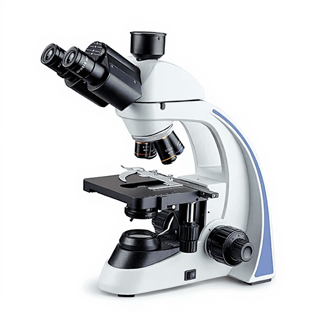
40X~1000X Digital Trinocular Biological Microscope
Research-grade trinocular biological microscope with integrated digital imaging, offering 40X-1000X magnification and 4K HDMI output for specimen documentation and analysis.

Louise Corscadden, PhD
Director of Science · ConductScience
Ask Louise about 40X~1000X Digital Trinocular Biological Microscope fit, setup, configuration, or quote prep.
Already working with us? Sign in to connect this with My Scientist.
Key Specifications
Full details →- Model fit
- Configured during quote
- SKU family
- MCX-0001
- Sizing
- 3.5 x 27.0 x 18.0 cm
- Ordering
- Online checkout and quote request available
- Category
- Microscopy
- Build notes
- Confirm accessories, station layout, and support needs before purchase
The 40X~1000X Digital Trinocular Biological Microscope is a research-grade optical instrument designed for detailed examination of biological specimens. This system combines traditional bright-field microscopy with integrated digital imaging capabilities, featuring a trinocular head configuration that accommodates simultaneous visual observation and digital documentation. The microscope incorporates a quadruple nosepiece with achromatic objectives spanning 4X to 100X magnifications, paired with WF10X eyepieces to achieve total magnifications from 40X to 1000X.
The integrated SONY IMX CMOS sensor provides high-resolution digital output via HDMI interface, supporting both 1080p at 60 FPS and 4K resolution at 30 FPS. The mechanical stage system offers precise specimen positioning with 70mm x 50mm travel range, while the Abbe condenser with N.A. 1.25 ensures optimal illumination control. LED illumination with brightness adjustment provides consistent lighting conditions for extended observation sessions.
How It Works
The microscope operates on the principle of bright-field transmitted light microscopy, where illumination from the LED source passes through the specimen and is collected by the objective lens system. The Abbe condenser with N.A. 1.25 focuses light onto the specimen plane, while the iris diaphragm controls illumination angle and contrast. Light transmitted through the specimen is collected by the achromatic objective lenses, which are corrected for chromatic and spherical aberrations to provide clear, high-contrast images.
The trinocular head configuration splits the light path, directing a portion to the binocular eyepieces for visual observation while simultaneously routing light to the integrated SONY IMX CMOS sensor. This 1/1.8-inch sensor with 2.0 μm pixel size captures high-resolution digital images that are processed and output via HDMI interface. The coaxial focusing system provides precise specimen positioning with 2 μm fine focus increments, enabling detailed examination at high magnifications up to 1000X with the oil immersion objective.
Features & Benefits
Automation Level
- semi-automated
Product Type
- Microscope
Microscope Type
- Biological / Compound
Viewing Head
- Trinocular
Application
- Clinical Research
- Life Science
Skill Level
- Entry Level
Brand
- ConductScience
Research Domain
- Cell Biology
- Clinical Diagnostics
- Developmental Biology
- Histopathology
- Materials Science
- Microbiology
Weight
- 0.2 kg
Dimensions
- L: 3.5 mm
- W: 27.0 mm
- H: 18.0 mm
| Feature | This Product | Typical Alternative | Advantage |
|---|---|---|---|
| Digital Output Resolution | 4K (3840×2160) at 30 FPS and 1080p at 60 FPS via HDMI | Entry-level models often limited to 1080p or require separate USB cameras with lower frame rates | Higher resolution enables detailed documentation and analysis of fine cellular structures. |
| Sensor Technology | SONY IMX CMOS with 2.0 μm pixel size on 1/1.8" format | Basic systems may use smaller sensors or older CCD technology with larger pixel sizes | Advanced CMOS technology provides better sensitivity and lower noise for clearer digital images. |
| Objective Configuration | Four achromatic objectives (4X/0.1, 10X/0.25, 40X/0.65, 100X/1.25) in quadruple nosepiece | Budget models may include fewer objectives or lower numerical apertures | Complete magnification range with high N.A. objectives supports diverse specimen types and research applications. |
| Focus Precision | Coaxial coarse/fine focus with 2 μm minimum division | Basic systems often have 5-10 μm focus increments or separate focus controls | Ultra-fine focus control essential for high-magnification work and critical focusing of thin specimens. |
| Stage System | Double-layer mechanical stage with 70mm × 50mm travel range | Single-layer stages or limited travel ranges common in entry-level models | Enhanced stability and larger scanning area improves precision for systematic specimen examination. |
| Interface Compatibility | C-mount lens interface with HDMI digital output | Proprietary connections or USB-only output limits accessory compatibility | Standard C-mount interface ensures compatibility with various imaging accessories and professional documentation systems. |
This system combines research-grade optics with integrated digital imaging capabilities, offering both 4K output resolution and ultra-fine focus control. The SONY CMOS sensor and comprehensive objective set provide professional documentation capabilities typically requiring separate camera systems.
Practical Tips
Verify magnification accuracy using a stage micrometer at each objective power, particularly after transport or major adjustments.
Why: Ensures measurement accuracy and confirms proper optical alignment across all magnifications.
Clean objectives with appropriate lens paper and solvents, working from center outward in circular motions.
Why: Proper cleaning prevents optical degradation and maintains image quality, especially critical for high N.A. objectives.
Allow LED illumination to warm up for 10-15 minutes before critical imaging work to achieve stable color temperature.
Why: Temperature stabilization ensures consistent illumination and color balance for quantitative imaging applications.
If digital and visual images appear differently focused, check C-mount adapter parfocality and sensor position.
Why: Parfocal alignment between visual and digital paths is essential for consistent documentation without refocusing.
Use the highest frame rate (60 FPS at 1080p) for live observation and 4K mode for final documentation images.
Why: Balances real-time responsiveness during specimen search with maximum resolution for permanent records.
Always use immersion oil sparingly and clean objectives immediately after use to prevent oil migration to lower power objectives.
Why: Prevents contamination of dry objectives and maintains optical performance across the entire objective set.
Adjust condenser height and iris diaphragm for each objective change to optimize contrast and resolution.
Why: Proper Kohler illumination setup maximizes image quality and ensures optimal performance of each objective's numerical aperture.
Store the microscope with lowest power objective in position and stage lowered to protect objectives from accidental contact.
Why: Reduces risk of objective damage and maintains mechanical system alignment during storage periods.
Setup Guide
What’s in the Box
- Digital trinocular biological microscope
- WF10X eyepieces (pair)
- Achromatic objectives: 4X/0.1, 10X/0.25, 40X/0.65, 100X/1.25
- HDMI cable
- Power adapter and cord
- Immersion oil (typical)
- Dust cover
- User manual and software disc (typical)
- Calibration slides (typical)
Warranty
ConductScience provides a standard one-year manufacturer warranty covering defects in materials and workmanship, with technical support available for setup assistance and operational guidance.
Compliance
References
Background reading relevant to this product:
What is the resolution limit at maximum magnification?
At 1000X magnification using the 100X/1.25 N.A. oil immersion objective, theoretical resolution approaches 0.22 μm, though practical resolution depends on specimen preparation and illumination conditions. Consult product datasheet for measured resolution specifications.
Can the digital output be used for time-lapse imaging?
The HDMI output supports continuous 60 FPS at 1080p resolution, making it suitable for real-time documentation. For extended time-lapse applications, external recording equipment would be required as internal storage capacity is not specified.
Is the system compatible with fluorescence applications?
This is a bright-field microscope with LED transmitted illumination. Fluorescence capability would require additional filter sets and appropriate excitation sources not included with the base system.
What specimen thickness can be accommodated?
The mechanical stage and focusing system can accommodate standard microscopy preparations including slides up to 1.2mm thickness. Working distance varies by objective, with the 100X oil immersion having the shortest working distance.
How does the digital image quality compare to eyepiece observation?
The SONY IMX CMOS sensor with 2.0 μm pixels provides high-resolution digital capture, though direct comparison depends on monitor quality and viewing conditions. Both paths share the same objective optics for consistency.
What maintenance is required for the LED illumination?
LED illumination typically requires minimal maintenance compared to traditional halogen systems. Expected lifespan and replacement procedures should be confirmed in the product documentation.
Have a question about this product?
Have a question? Just ask.
Send it over and we'll email you a personalized answer — no call, no scheduling.
Prefer to talk it through?
Accessories
Enhance your setup with compatible accessories

Frequently Bought Together


Upgrade Options
Related Products