Integrated Horizontal Electrophoresis System
Compact horizontal electrophoresis system with integrated blue light transilluminator for rapid agarose gel separation of DNA and RNA samples, featuring four gel sizes and comprehensive comb sets.
| Instrument Type | Electrophoresis |
| Application Area | Protein Analysis |
| Automation Level | semi-automated |
| gel_pore_size | 100-500nm diameter |
| number_of_gel_sizes | 4 different sizes |
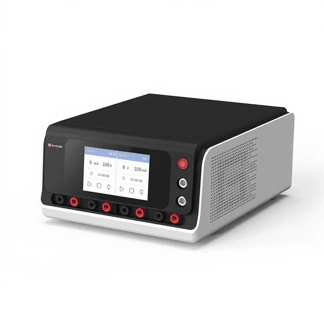
| power_supply_included | SPW-6S electrophoresis power supply |
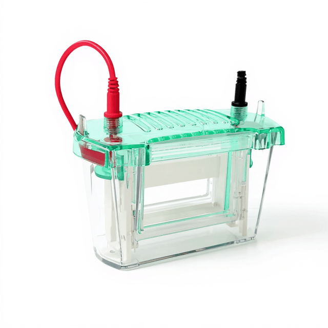
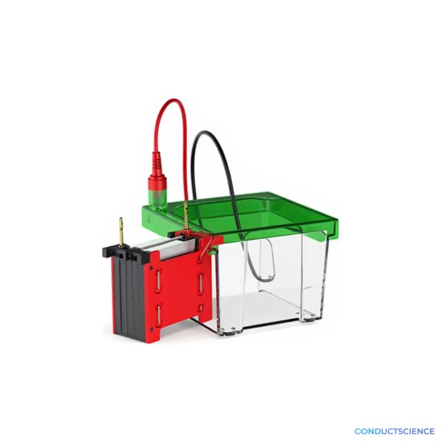
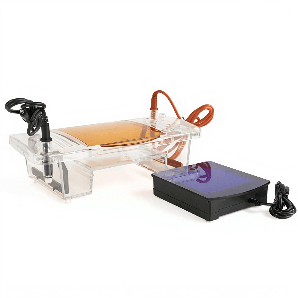
The Integrated Horizontal Electrophoresis System (SB-SVL-2) is a compact, multi-format gel electrophoresis apparatus designed for rapid agarose gel separation of DNA and RNA samples. The system features an integrated gel maker capable of producing four different gel sizes and includes a built-in blue light transilluminator operating at 470nm for nucleic acid visualization. With a 260mL buffer capacity and dimensions of 310 x 150 x 120mm, this system provides flexibility for various sample throughput requirements in molecular biology laboratories.
The system includes multiple gel tray configurations (120x120mm, 120x60mm, 60x120mm, and 60x60mm) and comprehensive comb sets with 1.0mm, 1.5mm, and 2.0mm thickness options, accommodating different sample volumes and resolution requirements. The integrated design eliminates the need for separate transillumination equipment, streamlining workflow and reducing bench space requirements for routine nucleic acid analysis.
How It Works
Horizontal gel electrophoresis separates nucleic acids based on size and charge through migration in an electric field. DNA and RNA samples, mixed with loading dye, are loaded into wells formed by combs in the agarose gel matrix. When voltage is applied across the gel via buffer-filled chambers, negatively charged nucleic acids migrate toward the positive electrode, with smaller fragments moving faster through the gel pores than larger fragments.
The agarose gel concentration determines separation resolution, with the gel pore size of 100-500nm diameter providing optimal separation for most DNA fragment ranges. The system's 470nm blue light transilluminator excites fluorescent nucleic acid stains, making DNA and RNA bands visible for documentation and analysis. The integrated design eliminates the need to transfer gels between electrophoresis and visualization equipment.
Features & Benefits
Instrument Type
- Electrophoresis
Application Area
- Protein Analysis
Automation Level
- semi-automated
gel_pore_size
- 100-500nm diameter
number_of_gel_sizes
- 4 different sizes
power_supply_included
- SPW-6S electrophoresis power supply
transilluminator_type
- blue light transilluminator
buffer_chambers
- 2
gel_type_compatibility
- agarose gel
gel_tray_sizes
- 120mm x 120mm, 120mm x 60mm, 60mm x 120mm, 60mm x 60mm
sample_types
- DNA and RNA samples, protein complexes
gel_making_capacity
- up to 2 pieces of 60mm x 60mm gel simultaneously
Brand
- Servicebio
Material
- Acrylic
Research Domain
- Analytical Chemistry
- Clinical Diagnostics
- Environmental Monitoring
- Microbiology
- Molecular Biology
- Pharmaceutical QC
Weight
- 2.2 lbs
Dimensions
- L: 12.2 in
- W: 5.91 in
- H: 4.72 in
Comparison Guide
| Feature | This Product | Typical Alternative | Advantage |
|---|---|---|---|
| Gel Format Options | Four gel sizes from 60x60mm to 120x120mm with unified gel maker | Many systems offer single fixed gel size or require separate casting equipment | Provides flexibility to match gel size to sample throughput without requiring multiple systems or additional equipment. |
| Integrated Transillumination | Built-in 470nm blue light transilluminator included | Most basic systems require separate UV or blue light boxes | Eliminates gel transfer steps and reduces equipment costs while maintaining compact bench footprint. |
| Buffer Capacity | 260mL total buffer volume with dual chambers | Entry-level models often have smaller buffer volumes requiring more frequent changes | Supports extended run times and maintains stable current distribution for consistent separation results. |
| Comb Selection | Multiple thickness options (1.0mm, 1.5mm, 2.0mm) with varying tooth counts | Basic systems typically include limited comb options | Optimizes well volume and sample loading capacity for different concentration ranges and applications. |
| System Weight | 1kg total weight with integrated components | Separate transillumination equipment adds significant weight and complexity | Maintains portability for shared laboratory use while providing complete functionality in single integrated unit. |
| Construction Material | Acrylic construction with chemical resistance | Varies by model, with some using less durable plastics | Provides long-term compatibility with electrophoresis buffers and standard laboratory cleaning protocols. |
This integrated system combines horizontal electrophoresis with blue light transillumination in a compact 1kg unit, offering four gel format options and comprehensive comb selection. The 260mL buffer capacity and unified gel maker provide operational flexibility while the integrated design streamlines nucleic acid analysis workflows compared to systems requiring separate visualization equipment.
Practical Tips
Use DNA ladder standards of known molecular weights with each gel run to verify separation performance and enable accurate fragment size determination.
Why: Molecular weight standards provide reference points for fragment size calculation and detect any changes in separation efficiency.
Clean gel trays and combs immediately after use with warm water and mild detergent to prevent agarose buildup that can affect well formation.
Why: Residual agarose can create uneven well bottoms and affect sample loading consistency in subsequent runs.
Allow adequate time for gel polymerization before removing combs and ensure buffer levels completely cover the gel surface during runs.
Why: Proper gel setting prevents well collapse during loading while adequate buffer coverage ensures uniform current distribution.
If bands appear distorted or smeared, check that gel concentration matches fragment size range and verify even buffer distribution in both chambers.
Why: Gel concentration mismatches or uneven current flow are common causes of poor separation resolution and band quality.
Use consistent gel thickness and agarose concentration between runs when comparing fragment sizes across different experiments.
Why: Standardized gel parameters ensure reproducible migration patterns and accurate molecular weight comparisons.
Always disconnect power supply before handling gels or accessing the electrophoresis chamber to prevent electrical shock hazards.
Why: Electrophoresis buffers conduct electricity and contact with energized systems can cause serious injury.
Pre-cool the blue light transilluminator before gel placement to minimize heat-induced gel distortion during visualization and documentation.
Why: Heat from transillumination can cause gel expansion that distorts band positions and affects size determination accuracy.
Inspect electrode connections regularly for corrosion or buffer salt buildup that can affect current flow and separation consistency.
Why: Electrode degradation creates uneven current distribution leading to irregular migration patterns and reduced separation quality.
Setup Guide
What’s in the Box
- Horizontal electrophoresis apparatus (1 set)
- Gel maker (1)
- 1.0mm 25-tooth/11-tooth sample combs (4)
- 1.5mm 13-tooth/6-tooth sample comb (1)
- 2.0mm 3-tooth/2-tooth sample comb (1)
- 60mm x 60mm gel trays (2)
- 60mm x 120mm gel tray (1)
- 120mm x 60mm gel tray (1)
- 120mm x 120mm gel tray (1)
- Blue light transilluminator (1 set)
- User manual and documentation (typical)
- SPW-6S electrophoresis power supply
Warranty
ConductScience provides a standard one-year manufacturer warranty covering defects in materials and workmanship, with technical support available for troubleshooting and application guidance.
Compliance
What gel concentrations are recommended for different DNA fragment size ranges?
The 100-500nm pore size range accommodates standard agarose concentrations from 0.8-2.0%, with lower concentrations for larger fragments (>1kb) and higher concentrations for small fragments (<500bp). Consult gel preparation protocols for specific concentration recommendations.
Can this system accommodate different buffer systems besides standard TAE or TBE?
The 260mL buffer capacity and acrylic construction are compatible with standard electrophoresis buffers including TAE, TBE, and Bis-Tris systems. Verify buffer compatibility with acrylic materials for specialized buffer formulations.
What is the maximum voltage recommended for each gel size?
Voltage recommendations depend on gel size and desired separation time. Consult the SPW-6S power supply specifications and gel electrophoresis protocols for optimal voltage settings per gel configuration.
How does the integrated blue light transilluminator compare to UV systems for sensitivity?
The 470nm blue light system requires nucleic acid stains optimized for blue light excitation and generally provides safer operation than UV systems. Sensitivity depends on the specific fluorescent dye used with the samples.
What maintenance is required for the gel maker and electrophoresis chambers?
Regular cleaning with mild detergent removes buffer salts and residual agarose. The acrylic construction allows standard laboratory cleaning protocols, avoiding harsh solvents that may cause stress cracking.
Can multiple gels be prepared simultaneously with the integrated gel maker?
The gel maker can accommodate up to 2 pieces of 60mm x 60mm gel simultaneously according to the specifications. Larger gel formats require individual preparation.
What documentation capabilities are available with the blue light transilluminator?
The system includes blue light visualization at 470nm wavelength. Documentation capabilities depend on external imaging equipment compatibility. Consult product datasheet for specific imaging system requirements.
How does this system compare to vertical electrophoresis for protein analysis applications?
This horizontal system is optimized for nucleic acid analysis using agarose gels, while vertical systems are typically used for protein analysis with polyacrylamide gels. The application determines the appropriate electrophoresis format.
Have a question about this product?
Accessories
Enhance your setup with compatible accessories