ConductVision Organoid Monitor
Published research on organoid imaging
Read which published studies support a pilot's measurements, what a camera cannot measure, and how a pilot checks ConductVision against organoids measured by hand.
OrganoID's published accuracy
OrganoID compared with organoids measured by hand
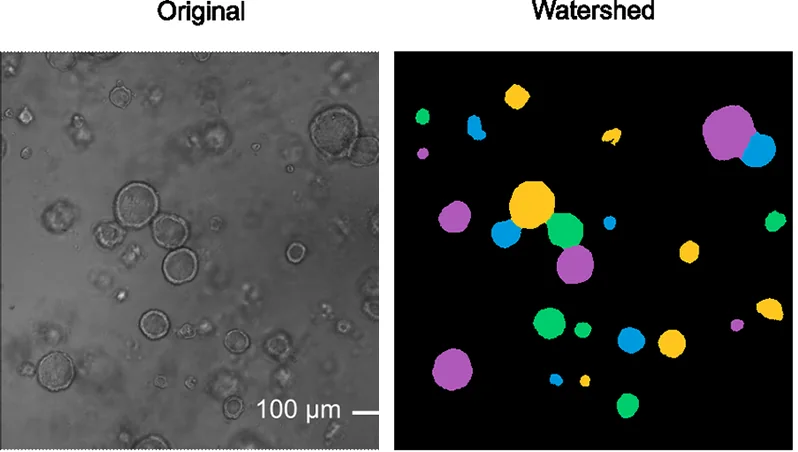
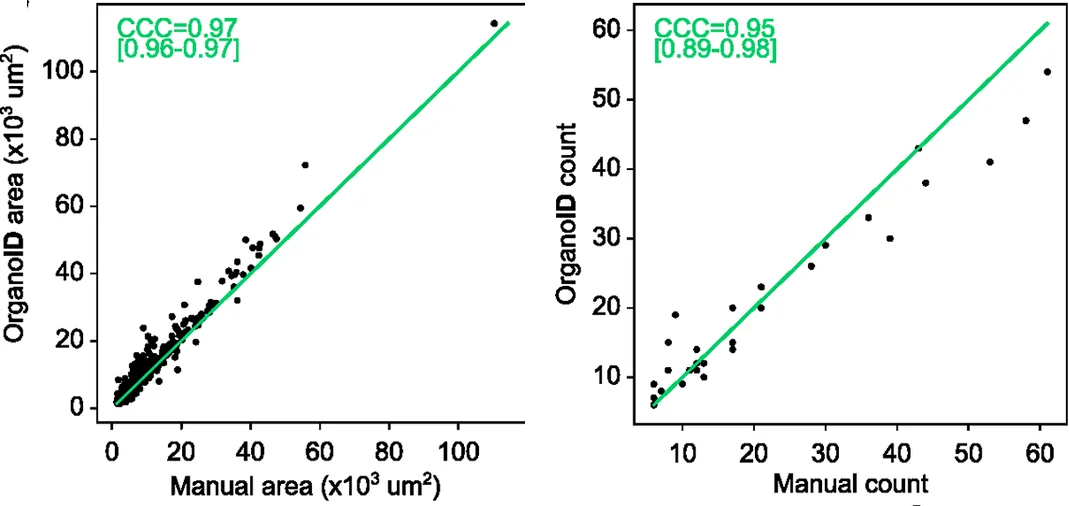
OrganoID, a published deep learning tool, finds, labels and tracks single organoids in brightfield and phase-contrast microscope images.1 Its organoid counts and areas agreed closely with measurements made by hand on the same images.1 Over a four-day time-lapse, it correctly matched over 89% of the organoids it identified from one time step to the next.1
Other studies
What other published tools measured
Other groups reported these results from their own tools and imaging systems.
| Measure | Published result |
|---|---|
| Count and area from brightfield alone | OrBITS counts, mean areas and total areas from brightfield images correlated with the same measures from Hoechst nuclear staining, with correlation coefficients of 0.98, 0.93 and 0.95.5 |
| Many organoids at once | OrganoSeg profiled 5,167 breast cancer spheroids and 5,743 organoids from colorectal cancer patients' normal and tumor tissue in brightfield images.3 |
| Intestinal organoid size | OrgaQuant located human intestinal organoids in brightfield images and measured their size distribution.2 |
Pilot check
How a pilot checks the camera
In a pilot, a ConductVision engineer measures sample organoids by hand on microscope images of the same wells. The pilot reports how closely the camera's measurements match those hand measurements before any result is used.
Beyond the camera
What a camera cannot measure
- Individual cells and nuclei
- Cell division (mitosis)
- Structures inside cells, including mitochondria
- Individual neurons and fine neurites
- How single cells move inside an organoid
- Cell polarity
- Protein expression and molecular markers
- Fine vascular networks
- Membrane potential and calcium signaling
- Gene expression in single cells
- Fine epithelial layers
- Cell layout inside crypts
- Nephron structures
Reading list
Reading list
Measuring organoids from images
- OrganoID tracks single organoids in brightfield and phase-contrast images.Matthews JM, Schuster B, Kashaf SS, et al. (2022). OrganoID: A versatile deep learning platform for tracking and analysis of single-organoid dynamics. PLOS Computational Biology.
- OrgaQuant locates and sizes intestinal organoids in brightfield images.Kassis T, Hernandez-Gordillo V, Langer R, et al. (2019). OrgaQuant: Human Intestinal Organoid Localization and Quantification Using Deep Convolutional Neural Networks. Scientific Reports.
- OrganoSeg measures size and shape across thousands of organoids and spheroids.Borten MA, Bajikar SS, Sasaki N, et al. (2018). Automated brightfield morphometry of 3D organoid populations by OrganoSeg. Scientific Reports.
- MOrgAna analyzes organoid images from stereoscopes to confocal systems.Gritti N, Lim JL, Anlaş K, et al. (2021). MOrgAna: accessible quantitative analysis of organoids with machine learning. Development.
Monitoring over time
- OrBITS follows organoids through drug screens from brightfield images alone.Deben C, De La Hoz EC, Compte ML, et al. (2023). OrBITS: label-free and time-lapse monitoring of patient derived organoids for advanced drug screening. Cellular Oncology.
- Days of live imaging show lumen cycles, migration, rotation and fusion.Hof L, Moreth T, Koch M, et al. (2021). Long-term live imaging and multiscale analysis identify heterogeneity and core principles of epithelial organoid morphogenesis. BMC Biology.
- Brightfield images follow individual tumor organoids through drug responses without dyes.Spiller ER, Ung N, Kim S, et al. (2021). Imaging-Based Machine Learning Analysis of Patient-Derived Tumor Organoid Drug Response. Frontiers in Oncology.
Responses across organoids
- Drug effects differ between organoids and flat cultures of the same cells.Jabs J, Zickgraf FM, Park J, et al. (2017). Screening drug effects in patient-derived cancer cells links organoid responses to genome alterations. Molecular Systems Biology.
Imaging inside the incubator
- The Picroscope images 24-well plates over time from low-cost parts.Ly VT, Baudin PV, Pansodtee P, et al. (2021). Picroscope: low-cost system for simultaneous longitudinal biological imaging. Communications Biology.
Plan an organoid monitoring pilot
Tell us which measure you want checked on your own plates.