ConductVision Organoid Monitor
Cameras, data files and quality checks
A pilot sets fixed cameras with macro lenses and controlled lighting inside your incubator or in a chamber beside it, and delivers every measurement as files you can analyze.
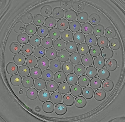
Camera specification
The cameras a pilot uses
ConductScience supplies pilot cameras that meet every requirement below.
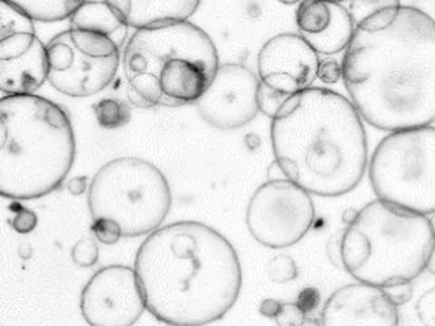
Image scale
- 2 µm or less per pixel
- A lens that resolves group 7, element 1 of a USAF 1951 resolution chart, whose lines are about 4 µm wide, through a culture plate, at the center and edges of the image
- Scale calibrated at every focal plane, so a change in depth does not read as growth
The Picroscope, an incubator camera system from another group, resolved the same chart element across a field about 5 mm wide.1
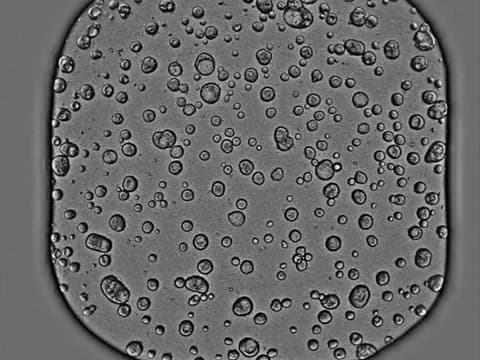
Field of view
- Each whole well, in one image or in tiles joined into one
- Enough cameras, or camera movement, to meet the timing below
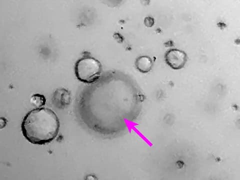
Focus
- Motorized focus
- Focal planes close enough that every organoid is sharp in at least one, through the full height of the gel
- Each organoid measured in the plane where it is sharpest
Timing
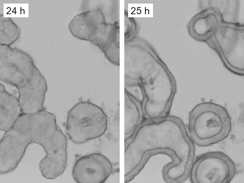
- A full stack of every well every 5 to 15 minutes
- An image a minute of a well while an event lasts, without delaying the other wells
- Clocks synchronized across cameras
Light
- Brightfield, with white LED light passing through the well to the camera
- Lights on only while a picture is taken
- The same light and exposure for the whole study
- Each image normalized to its own background
- Visible light, with infrared lighting as an option
Incubator
- Rated for 37 °C and high humidity for a whole study, inside an incubator or in a chamber beside it that keeps the same conditions
- Images with the plate lid on
- A heated window or lens that keeps condensation off the image
- Warms the plate by less than 0.5 °C, with the plate temperature logged and an alert if it drifts
- Shuts itself down if it overheats
- A plate holder that puts each well back in place
The Picroscope ran inside an incubator, with a relay board that can shut the system down if it overheats.1
Options
- More cameras
- A high-frame-rate camera for cardiac clips, at more than 70 frames per second
- Telecentric lenses, which keep an organoid's measured size the same at different depths
- Motorized plate movement
- Lighting from several angles
The authors of MUSCLEMOTION, a published tool that measures contraction from video, recommend more than 70 frames per second to time each contraction.2
Scale
One dish or a whole plate
A pilot can run one camera on one dish, or enough cameras to cover a whole plate. The Picroscope, a low-cost system that another group built mostly from off-the-shelf and 3D-printed parts, has already imaged standard 24-well plates over time, including 3D tissue cultures inside an incubator.1
Recording
Three ways a pilot records
- 1
Continuous imaging
A stack of focal planes every 5 to 15 minutes, to measure growth, shape and movement.
- 2
High-speed clips
With the high-frame-rate camera option, short clips in one focal plane at more than 70 frames per second, for beating cardiac organoids.
- 3
Event-triggered recording
When an event starts, such as an organoid fragmenting, the camera images that well more often in one focal plane, then returns to its routine rate to save storage.
Quality control
Image problems are flagged before any number is reported
Measurements from a flagged frame stay in your data files with a flag, and the summaries and report leave them out.
Environment
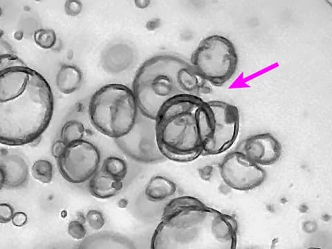
- Condensation
- Obstruction
Position
- Camera movement
- Plate movement
- Field drift
Records
- Missing frames
- Timestamp checks
Outputs
What you get back
Outlines and IDs
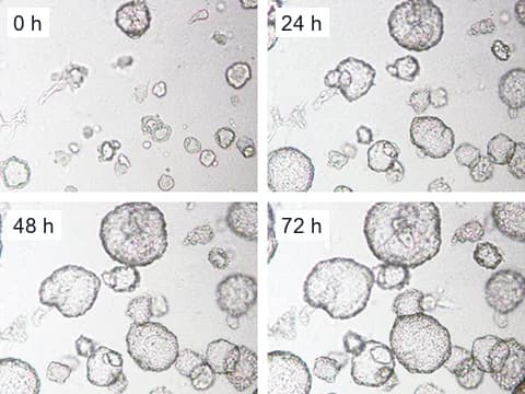
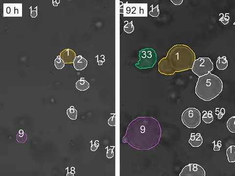
- Organoid outlines
- Organoid IDs
- Tracks
- Fusion and fragmentation records
Measurements
- Time series as CSV, JSON or Parquet
- Columns for timestamp, plate, well, organoid, area, growth rate, circularity and movement
Events
- A machine-readable event log
Report
- Growth curves
- Treatment effects
- The spread across organoids
- Unusual events
- Image-quality issues
- Representative images and clips
Questions labs ask
How do I know it will work with my plates?
A pilot checks your plate format, well size and culture method during setup. The setup record lists the wells each camera covers, the gel height and the number of focal planes. It then reports which measurements your images are sharp enough for.
Does it image fluorescence?
Not in the standard setup, which images in visible light.
References
- Ly VT, Baudin PV, Pansodtee P, et al. (2021). Picroscope: low-cost system for simultaneous longitudinal biological imaging. Communications Biology.
- Sala L, van Meer BJ, Tertoolen LG, et al. (2018). MUSCLEMOTION: A Versatile Open Software Tool to Quantify Cardiomyocyte and Cardiac Muscle Contraction In Vitro and In Vivo. Circulation Research.
- Deben C, De La Hoz EC, Compte ML, et al. (2023). OrBITS: label-free and time-lapse monitoring of patient derived organoids for advanced drug screening. Cellular Oncology.
- Hof L, Moreth T, Koch M, et al. (2021). Long-term live imaging and multiscale analysis identify heterogeneity and core principles of epithelial organoid morphogenesis. BMC Biology.
- Kassis T, Hernandez-Gordillo V, Langer R, et al. (2019). OrgaQuant: Human Intestinal Organoid Localization and Quantification Using Deep Convolutional Neural Networks. Scientific Reports.
- Lefferts JW, Kroes S, Smith MB, et al. (2024). OrgaSegment: deep-learning based organoid segmentation to quantify CFTR dependent fluid secretion. Communications Biology.
- Rohe J, Ashraf APK, Kehl M, et al. (2026). Modelling kidney cystogenesis using human kidney tubuloid cultures. BMC Molecular and Cell Biology.
- Matthews JM, Schuster B, Kashaf SS, et al. (2022). OrganoID: A versatile deep learning platform for tracking and analysis of single-organoid dynamics. PLOS Computational Biology.
Plan the camera setup for your plates
Tell us your plate format, your incubator and the first question you want answered.