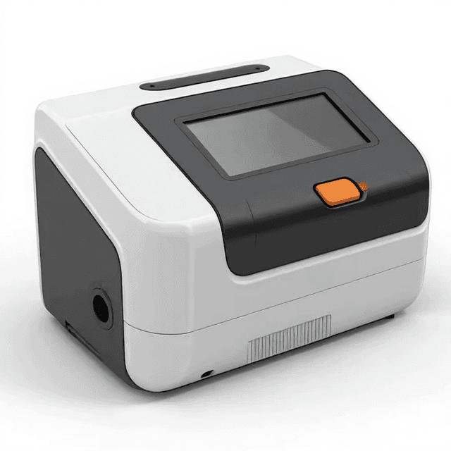
Automated Fluorescence Cell Counter
Our Automated Cell Counter is an advanced laboratory instrument designed for rapid and precise quantification of cells in a given sample. Equipped with state-of-the-art image processing technology, it automates the cell counting process, eliminati...
| counting_time_brightfield | 9 seconds |
| counting_area | 2.15 mm×1.62 mm |
| cell_size_range | 4-60 microns |
| concentration_range | 104-107 cells/mL |
| amplification_factor | 2.5×5.0 Megapixels |
| focusing_methods | manual focusing and autofocusing |
Introduction
An automated cell counter is an instrument for automatically counting living and/or dead cells. Researchers opt for automated cell counters compared to manual hemocytometers, which have been used to count cells for decades since they deliver faster and more accurate cell counts. An automated cell counter can deliver a count in less than 9 seconds in the bright field mode compared to the 5 minutes manual counting would take.
An automated cell counter can measure cell concentration before cell passage or evaluate the cell viability of a cultured cell line. Conduct Science’s automated cell counter allows easy operation of brightfield and fluorescence cell counts. It also has multiple data displays and storage, which furthers its convenience.
Specifications
| Parameters | Remarks |
|---|---|
| Amplification Factor | 2.5X, 5.0 Megapixel |
| Optional fluorescence application | (Ex :375/28nm ;Em:460/50nm)
(Ex :480/30nm ;Em:535/40nm) (Ex :540/25nm ;Em:620/60nm ) |
| Focus Method | Manual focusing & Autofocusing |
| Counting Area | 2.15 mm×1.62 mm |
| Cell Type | Cell lines, stem cells, primary cells, pollens, beer yeast |
| Cell Size Range | 4-60 μm (Optimal: 7-60 μm) |
| Cell Concentration Range | 10^4-10^7 cells/mL |
| Cell counting Time | Less than 9s |
| Historical Data Storage | 1000 counting reports and images at most |
| Optional fluorescence application | (Ex: 375/28nm; Em: 460/50nm) (Ex: 480/30nm; Em: 535/40nm) (Ex: 540/25nm; Em: 620/60nm) |
| Languages | Chinese and English |
| Dimensions | 212 mm(W)* 264 mm(H)* 165 mm (D |
| Weight | ~3.2 kg |
Features
High Accurate
- A high-definition imaging system and independently developed recognition algorithms technology enable C100 to count and analyze stem cells, primary cells, and cell lines accurately.
- Users can self-define and preset thresholds for cell size, brightness, and roundness to reduce counting errors.
- Parameters can be modified at any time to obtain the best cell imaging & more accurate counting results.
- Primary mouse macrophages and HEK293 cells were counted with a hemocytometer and automated counters resulting in the standard deviation of C100 was smaller than other counters and the CV value was less than 5%, while manual counting can reach more than 10%.
Fast
- In the bright field mode, a count can be completed in 9 seconds, while manual counting is usually up to 5 minutes.
- Fluorescence analysis operation is easy to use.
- A built-in dilution calculator can give a dilution solution quickly based on the existed counting result, facilitate the experiments and save a lot of time for operators.
- If each operator counts 10 cell samples (5pcs) per day, then 24.25 hours can be saved each month.
Convenient
- Multiple data display and storage, including cell images, diameter distribution histograms, count results, live and dead cells percentage.
- Convenient for storage and analysis, PNG, JEPG, CSV, PDF, etc.
- 1,000 historical counts can be stored in the device or exported via USB.
- 45° elevation angle, ergonomic design, reduced visual fatigue and cervical pressure.
- 7 inch high-definition touch screen with friendly interaction and comfortable operation.
Safe
- Disposable counting slides, no need to clean, reducing biological hazards risk.
Apparatus
The Automated Cell Counter weighs ~3.2 kg and measures 212 mm in width, 264 mm in height, and 165 mm in depth. It can count various cells such as stem cells, primary cells, cell lines, pollens, and beer yeast in less than 9 seconds. The counting area measures 2.15 mm×1.62 mm. It can accurately count cells with a size range of 4-60 microns and a concentration of 104-107 cells/mL. It has an amplification factor of 2.5×5.0 Megapixels. It also includes an option for fluorescence analysis. It allows both manual focusing and autofocusing methods. It has large historical data storage that can save up to 1000 counting reports and images. It can be used in either Chinese or English language settings.
Applications
Medical and research laboratories employ automated cell counters to assess the quantity and kinds of cells present in blood or urine samples and verify the viability of a cultivated cell line for research purposes.
The automated cell counter is highly efficient in counting deferential neutrophil counts, with moderate efficiency in counting lymphocytes and less efficiency in monocytes and eosinophils counting (von Konigslow, Renaud, Duffield, Higginson, & Kelton, 2019). The countermeasures cells of body fluids with precision, improved accuracy, and efficiency is extremely difficult with manual counters (Bourner et al., 2014).
Protocol
- Connect the Automated Cell Counter to your computer or laptop and turn it on
- Place the filled counting chamber on top of the stage
- Tap the Count button while concentrating on your cells
Strengths and Limitations
Strengths
Automated cell counting is quicker, less reliant on the user, and simple to use. The device has a large data storage, allowing 1000 counts to be stored. Moreover, it allows multiple data displays and storage with the data access allowed from any location, at any time.
It can count various types of cells that include cell lines, stem cells, beer yeast, primary cells, and pollens with the accuracy in size ranging from 4 microns to 60 microns. It only weighs 3.2 kg, which makes it easy to carry anywhere. In terms of safety, there is no hazardous biological damage as it comes with disposable slides.
Using it at a comfortable angle of 45 degrees reduces eye fatigue and spine pain. A built-in dilution calculator makes the work quicker and less tedious. Users can preset functions according to their needs; they can adjust cell size, roundness, and brightness which makes it more accurate. It can store data in different formats, i.e., PNG, PDF, CSV, and JPEG.
Limitations
Automated hematology analyzers may inadvertently exaggerate or deflate cell counts. They solely measure the volume and amount of particles. Certain analyzers, particularly impedance-based counters, may not distinguish between minute aggregates of platelets and nucleated red blood cells. Platelet clumping may be mistaken for leukocytes or erythrocytes, and nucleated red blood cells, especially lymphocytes, can be mistaken for leukocytes. Atypical cells that are big or unidentified, toxic immature neutrophils, and significantly reactive lymphocytes may also be misclassified.
Precautions
- Use the Counter within each model's ambient operating temperature, humidity, water, and oil exposure specifications.
- For each model, save the Counter with the set temperature range. Allow at least 3 hours for the Counter to come to room temperature if kept at a temperature below 10°C.
- Do not use the Counter in places with a lot of stress and vibration; long-term use in such settings might cause damage to the Counter.
- When switching a load, magnetic contactors produce a shock of 1,000 to 2,000 m/s2. Separate magnetic contactors from the Counter when mounting to DIN Track to avoid vibration and stress. Rubber with anti-vibration properties should be used.
- Organic solvents (benzene or paint thinner), strong alkalis, or strong acids should not be used since they will harm the Counter's exterior conditions.
- Keep the counter as far away from static electricity sources as possible.
- Condensation inside the Counter might cause malfunction or damage to the Counter's components.
- Depending on the operational climate, resin and rubber components (such as rubber packaging) may degrade, shrink, or harden (e.g., subjected to corrosive gases, ultraviolet light, or high temperatures). Periodic inspection and replacement are recommended.
Summary
- Automated cell counters are instruments for automatically counting living and dead cells in culture. They count suspension, adherent, and aggregated cells.
- They deliver faster, simpler, and more reliable cell counts at difficult-to-estimate concentrations than manual counters.
- Automatic cell counters may be self-contained (internal PC) or linked to an external computer.
- It allows the performance of image-based cell counts and also has an option for fluorescence analysis.
- It can accurately count cells with sizes ranging between 4-60 microns and concentrations ranging between 104-107 cells/mL.
References
Bourner, G., De la Salle, B., George, T., Tabe, Y., Baum, H., Culp, N., & Keng, T. B. (2014). ICSH guidelines for the verification and performance of automated cell counters for body fluids. International Journal of Laboratory Hematology, 36(6), 598-612. doi:10.1111/ijlh.12196
Green, R., & Wachsmann-Hogiu, S. (2015). Development, history, and future of automated cell counters. Clin Lab Med, 35(1), 1-10. doi:10.1016/j.cll.2014.11.003
von Konigslow, T. E., Renaud, D. L., Duffield, T. F., Higginson, V., & Kelton, D. F. (2019). Validation of an automated cell counter to determine leukocyte differential counts in neonatal Holstein calves. Journal of dairy science, 102(8), 7445–7452. https://doi.org/10.3168/jds.2019-16370
How It Works
The automated cell counter employs digital image analysis of cells suspended in a defined volume within disposable counting slides. The 2.5X optical magnification system with 5.0 megapixel resolution captures high-definition images of the 2.15 mm × 1.62 mm counting area. The instrument's image processing algorithms identify individual cells based on size parameters (4-60 μm diameter) and morphological characteristics, distinguishing viable cells from debris and cell aggregates.
Fluorescence capabilities utilize three excitation/emission wavelength combinations: 375/460 nm for DAPI-type nuclear stains, 480/535 nm for FITC-compatible dyes, and 540/620 nm for propidium iodide or similar viability indicators. The system integrates both manual and autofocusing mechanisms to optimize image clarity across different cell types and suspension densities. Cell concentration calculations account for the precise counting area volume and dilution factors when applicable.
Features & Benefits
Model
- C100
- C200FL
counting_time_brightfield
- 9 seconds
counting_area
- 2.15 mm×1.62 mm
cell_size_range
- 4-60 microns
concentration_range
- 104-107 cells/mL
amplification_factor
- 2.5×5.0 Megapixels
focusing_methods
- manual focusing and autofocusing
data_storage
- 1000 counting reports and images
language_settings
- Chinese or English
elevation_angle
- 45°
compatible_cell_types
- stem cells, primary cells, cell lines, pollens, beer yeast
fluorescence_analysis
- available
export_formats
- PNG, JPEG, CSV, PDF
export_method
- USB
consumables
- disposable counting slides
Automation Level
- fully-automated
Accuracy
- 2.5×5.0 Megapixels
- CV value less than 5%
Display Type
- 7 inch high-definition touch screen
Weight
- 3.2 kg
Dimensions
- 165 mm x 212 mm x 264 mm
Brand
- RWD
Research Domain
- Cancer Research
- Cell Biology
- Developmental Biology
- Immunology
- Microbiology
- Pharmaceutical QC
Weight
- 8.27 kg
Dimensions
- L: 21.2 mm
- W: 26.4 mm
- H: 16.5 mm
Comparison Guide
| Feature | This Product | Typical Alternative | Advantage |
|---|---|---|---|
| Analysis Speed | Less than 9 seconds per sample | Entry-level models often require 15-30 seconds | Increases laboratory throughput and reduces time spent on routine cell counting tasks. |
| Concentration Range | 10⁴ to 10⁷ cells/mL | Many systems offer narrower ranges requiring dilutions | Accommodates diverse sample types from dilute primary isolates to concentrated cultures without preprocessing. |
| Fluorescence Channels | Three excitation/emission wavelength combinations | Basic models typically offer single-channel fluorescence or brightfield only | Enables multi-parameter analysis including viability assessment and specific fluorescent marker detection. |
| Data Storage | 1000 counting reports with images | Limited storage capacity in lower-cost alternatives | Supports comprehensive experimental documentation and quality control review over extended periods. |
| Display Interface | 7-inch high-definition touchscreen | Smaller displays or basic LCD screens in budget models | Provides clear image visualization for immediate quality assessment and intuitive operation. |
| Cell Size Range | 4-60 micrometers (optimal 7-60 μm) | Some systems limited to narrower size ranges | Accommodates diverse cell types from small lymphocytes to larger adherent cells in suspension. |
This automated cell counter combines rapid analysis speed with comprehensive fluorescence capabilities and extensive data storage. The high-definition touchscreen interface and broad dynamic range support diverse laboratory workflows from routine culture maintenance to specialized research applications.
Practical Tips
Verify counting accuracy monthly using certified reference beads of known concentration within your typical working range.
Why: Regular validation ensures measurement traceability and identifies any drift in counting performance.
Clean the slide insertion area daily and optical components weekly using lint-free lens tissue and appropriate cleaning solutions.
Why: Optical cleanliness directly impacts image quality and counting accuracy, particularly for smaller cells near the detection threshold.
Prepare duplicate samples for critical experiments and compare results to assess measurement variability.
Why: Biological samples can exhibit heterogeneity that affects counting statistics, and duplicate analysis improves data confidence.
If counts appear consistently low, check for cell settling in the suspension and ensure gentle mixing before slide loading.
Why: Cell sedimentation can result in non-representative sampling and underestimation of true cell concentration.
Review captured images for debris, aggregates, or atypical morphology before accepting counting results.
Why: Visual verification of automated analysis helps identify sample preparation issues or instrument performance problems.
Handle fluorescent dyes according to manufacturer safety data sheets and dispose of counting slides as biohazardous waste.
Why: Fluorescent reagents may pose health hazards, and used slides contain biological material requiring proper disposal protocols.
Document cell passage numbers and culture conditions when storing counting data for experimental traceability.
Why: Cell behavior can change with passage number, and comprehensive documentation supports reproducible experimental protocols.
Adjust focus manually if autofocus struggles with samples containing high debris content or unusual cell morphology.
Why: Manual focus control provides better optimization for challenging samples that may confuse automated focusing algorithms.
Setup Guide
What’s in the Box
- Automated fluorescence cell counter main unit
- Power adapter and cable
- USB cable for data export
- User manual and quick start guide
- Disposable counting slides (typical)
- Calibration certificate (typical)
Compliance
References
Background reading relevant to this product:
Warranty & ConductCare
ConductScience provides a standard one-year manufacturer warranty covering defects in materials and workmanship, with technical support for operation and maintenance guidance.
What cell size range provides optimal counting accuracy?
The instrument performs optimally with cells sized 7-60 micrometers, though it can detect cells as small as 4 micrometers. Cells below 7 μm may be more susceptible to classification errors due to debris interference.
How does the fluorescence system distinguish live from dead cells?
The 540/620 nm excitation/emission channel accommodates propidium iodide staining for dead cell identification, while the 375/460 nm channel supports DAPI-type nuclear stains. Live/dead discrimination requires appropriate fluorescent dyes and staining protocols.
Can the system handle primary cells with irregular morphology?
Yes, the instrument accommodates primary cells, stem cells, and cell lines with diverse morphologies. The size discrimination range (4-60 μm) and image analysis algorithms account for shape variations common in primary isolates.
What sample preparation is required for accurate counting?
Samples should be single-cell suspensions without aggregates, debris minimized through filtration if necessary, and cell concentration within the 10⁴-10⁷ cells/mL range. Gentle mixing prevents sedimentation during loading.
How frequently should the instrument be cleaned and maintained?
Clean the slide insertion area daily with laboratory-grade alcohol. Monthly cleaning of optical components with lens tissue maintains image quality. Consult product datasheet for specific maintenance intervals.
What data export options are available for integration with LIMS?
The system exports data via USB in CSV format for spreadsheet analysis, PDF for reports, and PNG/JPEG for image documentation. The 1000-sample storage capacity supports batch data retrieval.
How does counting accuracy compare to manual hemocytometer methods?
Automated counting eliminates subjective operator variations and provides consistent results across users. The digital image analysis reduces counting fatigue effects common in manual methods, though specific accuracy metrics should be verified through validation studies.
Can the system differentiate between cell types in mixed populations?
The instrument provides size-based discrimination and fluorescence capabilities, but specific cell type identification requires appropriate fluorescent markers and validated staining protocols for the cell populations of interest.
Have a question about this product?
Accessories
Enhance your setup with compatible accessories