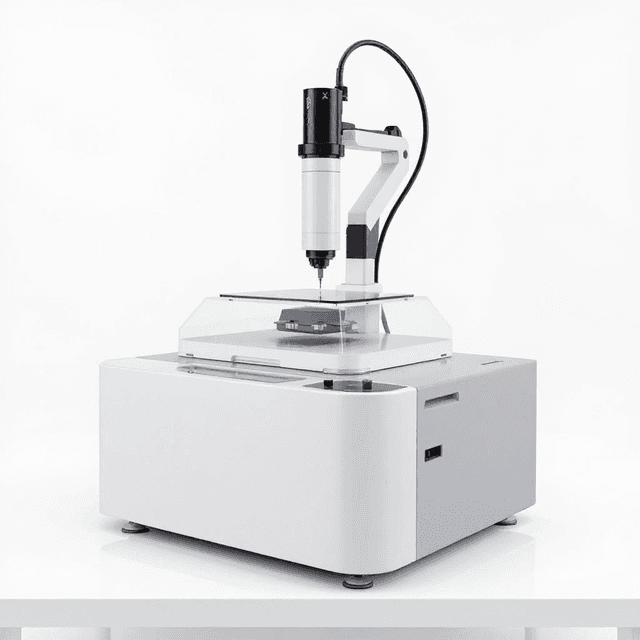
Automatic Gel Imaging and Analysis System
High-resolution gel documentation system with 5.03MP CCD camera, UV/white light sources, and motorized zoom lens for comprehensive electrophoresis analysis.
| Automation Level | semi-automated |
| Mode | BK-AG100 |
| Pixel | 2592*1944(5.03MP) |
| Exposure Time | 1ms~3000ms |
| Binning | 1*1 |
| OD | ≥4.8OD |
The Automatic Gel Imaging and Analysis System (BK-AG100) is a comprehensive documentation platform for electrophoresis gel analysis in molecular biology workflows. The system combines high-resolution CCD imaging (5.03 MP, 2592×1944 pixels) with both UV and white light illumination sources to capture protein bands, nucleic acids, and other biomolecules separated by gel electrophoresis. The motorized zoom lens (8-48mm, F1.2) provides precise focusing across a 21×21cm UV transillumination area.
This imaging system supports multiple detection modes including 302nm UV transillumination for nucleic acid visualization and LED-based epi-white illumination for protein gels and colorimetric assays. The optical density range of ≥4.8 OD enables detection of both faint and saturated bands within the same gel. Variable exposure times from 1ms to 3000ms accommodate different fluorophores and staining intensities, while the 590nm filter supports common fluorescent dyes with additional filters available for specialized applications.
How It Works
The system operates through CCD-based digital imaging of electrophoretically separated biomolecules. UV transillumination at 302nm excites DNA intercalating dyes (ethidium bromide, SYBR dyes) or RNA stains, while the high-sensitivity CCD sensor captures emitted fluorescence across the 2592×1944 pixel array. The motorized lens system automatically adjusts focal length from 8-48mm to optimize field of view and resolution for different gel sizes.
For protein analysis, the LED-based epi-white illumination system provides uniform top lighting for colorimetric detection of enzyme-linked substrates or direct protein stains. The optical system achieves ≥4.8 OD dynamic range through binning optimization and variable exposure control (1-3000ms), enabling simultaneous detection of low-abundance and high-abundance targets within the same gel.
Image acquisition occurs through direct digital capture without film processing, with the 590nm filter selectively transmitting fluorescence while blocking excitation wavelengths. The UV-to-white conversion sample plate enables direct comparison between fluorescent and colorimetric detection methods on the same gel.
Features & Benefits
Automation Level
- semi-automated
Mode
- BK-AG100
Pixel
- 2592*1944(5.03MP)
Exposure Time
- 1ms~3000ms
Binning
- 1*1
OD
- ≥4.8OD
Lens
- Motorized 8~48mm, F1.2
Trans-UV
- 302nm
Epi-White
- LED
Trans-White
- UV to white sample plate
Filters
- 590nm, Others for option
UV Area
- 21*21cm
Timing Off
- 1~60mins
Packing Size
- 460*475*725mm
Brand
- ConductScience
Research Domain
- Analytical Chemistry
- Cancer Research
- Cell Biology
- Developmental Biology
- Materials Science
- Microbiology
Power/Voltage
- 100~240V, 50/60Hz
Weight
- 31.0 kg
Dimensions
- L: 72.5 mm
- W: 46.0 mm
- H: 47.5 mm
Comparison Guide
| Feature | This Product | Typical Alternative | Advantage |
|---|---|---|---|
| Image Resolution | 5.03MP (2592×1944 pixels) with 1×1 binning | Entry-level systems often provide 1-3MP resolution | Higher pixel density enables detection of closely spaced bands and accurate molecular weight determination. |
| Exposure Time Range | 1ms to 3000ms variable control | Limited exposure ranges or fixed settings in basic models | Wide exposure latitude accommodates diverse fluorophore intensities and staining methods in a single platform. |
| Optical Density Range | ≥4.8 OD dynamic range | Standard systems typically offer 2-3 OD range | Extended dynamic range enables simultaneous detection of weak and strong signals without saturation. |
| Lens System | Motorized 8-48mm zoom, F1.2 aperture | Fixed focal length or manual adjustment systems | Automated optical control ensures reproducible magnification settings for quantitative analysis. |
| Illumination Sources | UV 302nm + LED epi-white dual system | Single illumination source in most basic models | Dual illumination supports both nucleic acid fluorescence and protein colorimetric detection without hardware changes. |
| UV Area Coverage | 21×21cm transillumination area | Smaller systems often limited to 15×15cm or less | Larger imaging area accommodates multiple gels simultaneously for high-throughput workflows. |
The BK-AG100 combines high-resolution CCD imaging with dual illumination sources and motorized optics for comprehensive gel documentation. The extended dynamic range and variable exposure control provide quantitative analysis capabilities suitable for research publication requirements.
Practical Tips
Use molecular weight standards in each gel run and verify consistent band migration patterns across the 21×21cm imaging area to maintain quantitative accuracy.
Why: Spatial calibration ensures accurate molecular weight determination and enables comparison between different gel positions.
Clean the UV transillumination surface weekly with ethanol to remove gel residue and maintain uniform light transmission across the imaging area.
Why: Surface contamination creates uneven illumination that affects band quantification and image quality.
Allow 15-minute UV lamp warm-up before critical imaging sessions and verify exposure settings with test samples to ensure reproducible results.
Why: UV lamp output stabilization prevents exposure variability that can affect quantitative band analysis.
Use the automated timing-off function to minimize UV exposure duration while maintaining adequate signal-to-noise ratios for band detection.
Why: Extended UV exposure can cause DNA nicking and sample degradation that affects subsequent analysis steps.
If bands appear unevenly illuminated, check that gels are positioned flat against the transillumination surface and verify filter alignment.
Why: Air gaps between gel and UV surface create focusing artifacts that reduce image sharpness and quantification accuracy.
Always use the provided UV shields and avoid direct eye exposure to the 302nm transillumination source during gel handling procedures.
Why: UV radiation at 302nm wavelength can cause corneal damage and skin burns with prolonged exposure.
Save imaging parameters (exposure time, zoom level, filter settings) as presets for specific gel types to ensure consistent documentation protocols.
Why: Standardized imaging conditions enable accurate comparison of band intensities across multiple experiments and time points.
Periodically verify motorized lens positioning accuracy using precision test targets to maintain focus quality throughout the zoom range.
Why: Mechanical wear in motorized systems can introduce focusing errors that reduce image sharpness and affect band edge definition.
Setup Guide
What’s in the Box
- BK-AG100 imaging system with integrated CCD camera
- UV transilluminator base (302nm)
- LED epi-white illumination module
- Motorized zoom lens assembly (8-48mm, F1.2)
- 590nm emission filter
- UV-to-white conversion sample plate
- Power adapter (100-240V)
- USB communication cable
- Analysis software CD/download
- User manual and safety guidelines (typical)
- Calibration reference standards (typical)
Warranty
ConductScience provides a standard one-year manufacturer warranty covering imaging components and electronics, with technical support for software installation and basic troubleshooting. Extended service plans available for high-usage laboratory environments.
Compliance
What gel formats are compatible with the 21×21cm transillumination area?
The imaging area accommodates standard mini and midi gel formats including 10×8cm, 15×15cm, and custom sizes up to 21×21cm. Multiple mini gels can be imaged simultaneously for parallel sample comparison.
How does the 5.03MP resolution compare to film-based documentation?
The 2592×1944 pixel CCD provides digital resolution equivalent to high-quality film with immediate image availability and quantitative analysis capabilities. Dynamic range of ≥4.8 OD exceeds typical film sensitivity.
Can the system detect chemiluminescent signals for Western blot applications?
The CCD sensor can detect chemiluminescence, though specific sensitivity depends on substrate intensity. The extended exposure capability (up to 3000ms) supports weak signal detection, though consult product datasheet for quantum efficiency specifications.
What fluorophores are compatible with the 590nm filter?
The 590nm filter supports orange-red emission fluorophores including propidium iodide, ethidium bromide, and similar dyes. Additional filters available for SYBR Green (520nm), DAPI (460nm), and other specific fluorophore combinations.
How does motorized lens control improve reproducibility?
Automated focus and zoom adjustment eliminates manual positioning variability, ensuring consistent magnification and focal plane across multiple imaging sessions for quantitative band comparison.
What file formats does the analysis software support?
Consult product datasheet for specific supported formats. Most gel documentation systems provide TIFF, JPEG, and proprietary formats with integrated densitometry analysis capabilities.
Is the UV output sufficient for thick agarose gels?
The 302nm UV transillumination typically penetrates standard 0.8-2% agarose gels up to 5-8mm thickness. For thicker gels, longer exposure times (up to 3000ms) may be required for adequate fluorophore excitation.
How does this compare to smartphone-based gel imaging apps?
This system provides calibrated illumination, controlled exposure parameters, and quantitative analysis capabilities not available in smartphone applications. The motorized optics and standardized conditions ensure reproducible results for publication-quality documentation.