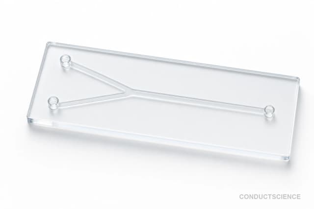
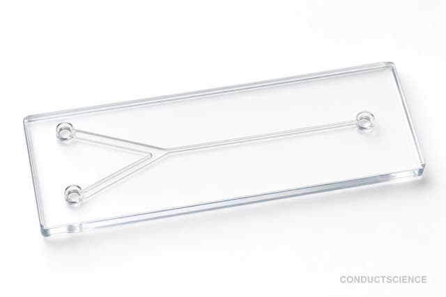
Standard PDMS Drug Screening Chip (200 um)
PDMS microfluidic chip with 200 x 200 μm channels designed for drug screening and efficacy testing applications.
Reusable chip — designed for multiple experimental runs. Compatible with standard microfluidic tubing: steel pins (0.7 mm ID / 1.0 mm OD) and silicone tubing (0.8 mm ID / 1.9 mm OD). Available in bulk packs (5‑, 10‑, and 25‑unit) for lab-scale and consumable workflows.

Louise Corscadden, PhD
Director of Science · ConductScience
Ask Louise about Standard PDMS Drug Screening Chip (200 um) fit, setup, configuration, or quote prep.
Already working with us? Sign in to connect this with My Scientist.
Key Specifications
Full details →- Model fit
- 3 selectable configurations
- SKU family
- WHM-0086
- Sizing
- 181.8 x 136.3 x 90.9 cm
- Ordering
- Online checkout and quote request available
- Category
- Drug Screening Chips
- Build notes
- Confirm accessories, station layout, and support needs before purchase
The Standard PDMS Drug Screening Chip (200 μm) is a microfluidic device manufactured from polydimethylsiloxane (PDMS) for pharmaceutical research applications. The chip features precisely engineered channels with dimensions of 200 x 200 μm, designed to facilitate controlled drug delivery and cellular response analysis in microscale environments. PDMS material provides optical transparency for real-time microscopy, biocompatibility for cell culture applications, and gas permeability for maintaining cellular viability during extended assays.
This microfluidic platform enables researchers to conduct drug screening experiments with reduced sample volumes, improved experimental control, and enhanced throughput compared to conventional well-plate methods. The standardized channel geometry ensures reproducible fluid dynamics and consistent drug concentration profiles across experimental replicates, making it suitable for quantitative pharmacological studies and efficacy testing protocols.
How It Works
Microfluidic drug screening operates on the principle of laminar flow within microscale channels. At the Reynolds numbers typical of microfluidic systems (Re < 1), viscous forces dominate over inertial forces, resulting in predictable, stratified flow patterns without turbulent mixing. This allows for precise spatial and temporal control of chemical concentrations within the device channels.
The PDMS chip utilizes pressure-driven flow to transport drug solutions through the 200 x 200 μm channels. Multiple inlet ports enable the creation of concentration gradients through controlled mixing of drug and buffer solutions. The channel dimensions are optimized to maintain laminar flow conditions while providing sufficient volume for cell culture and real-time observation.
Drug molecules diffuse across streamlines according to Fick's law, creating predictable concentration profiles that can be modeled and validated experimentally. The transparent PDMS material enables real-time fluorescence and bright-field microscopy for monitoring cellular responses, while the material's gas permeability maintains physiological oxygen and CO2 levels for cell viability during extended assays.
Features & Benefits
Pack Size
- 5-Pack
- 10-Pack
- 25-Pack
Weight
- 3.3 kg
Dimensions
- L: 181.8 mm
- W: 136.3 mm
- H: 90.9 mm
| Feature | This Product | Typical Alternative | Advantage |
|---|---|---|---|
| Channel Dimensions | 200 x 200 μm standardized geometry | Variable dimensions ranging from 50-500 μm depending on application | Provides optimal balance between cell culture volume and rapid drug transport kinetics. |
| Material Properties | PDMS construction with gas permeability | Glass or thermoplastic materials with limited permeability | Maintains cellular viability through oxygen transport while enabling optical transparency. |
| Fabrication Method | Soft lithography molding process | Varies by manufacturer and material choice | Enables rapid prototyping and cost-effective production for research applications. |
| Surface Treatment Options | Compatible with plasma treatment and protein coatings | Limited modification options for rigid materials | Allows customization of surface chemistry for specific cell types and experimental conditions. |
| Optical Compatibility | Transparent across visible spectrum | Material-dependent optical properties | Supports real-time microscopy and fluorescence imaging without optical interference. |
This device offers standardized channel geometry for reproducible drug screening experiments with PDMS material benefits including biocompatibility, optical transparency, and gas permeability. The 200 μm dimensions provide an optimal balance for cell culture and efficient drug transport kinetics.
| Model | SKU | Listed price | Status | Dimensions |
|---|---|---|---|---|
| 25-Pack | WHM-0086-25PK | Quote | Available | 181.8 x 136.3 x 90.9 cm |
| 10-Pack | WHM-0086-10PK | $1,490.00 | Available | 181.8 x 136.3 x 90.9 cm |
| 5-Pack | WHM-0086-5PK | Quote | Available | 181.8 x 136.3 x 90.9 cm |
Practical Tips
Degas PDMS devices under vacuum for 2-4 hours before first use to remove dissolved gases that can form bubbles during operation.
Why: Prevents bubble formation that can disrupt flow patterns and interfere with cell culture conditions.
Apply surface modifications immediately before cell loading to ensure optimal protein coating stability and cell adhesion.
Why: Time-dependent degradation of surface treatments can reduce coating effectiveness and cell attachment.
Store devices in sterile conditions with channels filled with PBS or culture medium to prevent drying and contamination.
Why: Dry channels can trap air and require extensive repriming, while contamination compromises experimental validity.
Establish baseline flow conditions and verify concentration gradients using fluorescent tracers before introducing cells or drugs.
Why: Ensures proper device function and validates theoretical predictions of concentration profiles.
Use consistent pressure sources and avoid rapid flow changes that can create shear stress variations affecting cellular responses.
Why: Mechanical stress can alter cellular behavior independent of drug effects, confounding experimental results.
Verify actual flow rates using microscopic particle tracking or direct volume measurement rather than relying solely on pump settings.
Why: Fluidic resistance variations between devices can cause flow rate deviations from programmed values.
Use appropriate chemical compatibility guidelines for PDMS when working with organic solvents or lipophilic compounds.
Why: Some chemicals can cause PDMS swelling or extraction of uncured oligomers that may affect experimental outcomes.
Setup Guide
What’s in the Box
- PDMS drug screening chip with 200 μm channels
- Product specification sheet (typical)
- Handling and storage instructions (typical)
Warranty
ConductScience provides a standard manufacturer warranty covering material defects and fabrication quality. Technical support is available for device handling, surface modification protocols, and experimental troubleshooting.
Compliance
What surface modifications are required for different cell types?
PDMS requires surface treatment for most cell culture applications. Use oxygen plasma treatment followed by protein coating (fibronectin, collagen) for adherent cells. Hydrophilic surface modifications improve wetting and reduce non-specific binding. Consult cell-specific protocols for optimal coating conditions.
How do I prevent air bubbles in the channels during operation?
Prime channels slowly with degassed buffer solutions. Use vacuum degassing of PDMS devices before first use to remove dissolved gases. Maintain consistent pressure and avoid rapid flow changes. Consider using bubble traps in the fluidic lines if necessary.
What flow rates are appropriate for drug screening applications?
Flow rates typically range from 0.1-10 μL/min depending on experimental requirements. Lower rates favor diffusion-dominated transport, while higher rates provide convection-dominated conditions. Calculate Péclet numbers to determine mass transport regimes for your specific application.
How long can cells survive in the microfluidic environment?
PDMS gas permeability supports cell viability for 24-48 hours under continuous flow conditions. For longer experiments, consider CO2-equilibrated media and controlled temperature environments. Monitor cell morphology and viability markers throughout extended cultures.
What microscopy setups are compatible with this device?
Standard inverted microscopes with long working distance objectives work well. PDMS thickness allows for both bright-field and fluorescence imaging. Consider refractive index matching for optimal optical performance. Device height may require objective correction for quantitative measurements.
How do I clean and reuse the chips?
PDMS devices can be cleaned with organic solvents, followed by oxygen plasma treatment for surface regeneration. However, protein adsorption and small molecule absorption may limit reusability for quantitative studies. Single-use is recommended for critical applications.
What drug concentrations can be achieved with this system?
Concentration ranges depend on inlet conditions and mixing ratios. Linear gradients from nanomolar to millimolar are achievable through controlled mixing. Consider drug solubility, stability, and PDMS absorption for accurate concentration delivery.
Have a question about this product?
Have a question? Just ask.
Send it over and we'll email you a personalized answer — no call, no scheduling.
Prefer to talk it through?