ConductVision Organoid Monitor
Brain and neural organoids, monitored 24/7
In a pilot, ConductVision measures the growth, outline and folds of each visible brain or neural organoid, from images taken every 5 to 15 minutes.
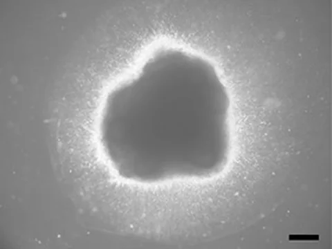
24/7 monitoring
What a pilot records for each brain or neural organoid
ConductVision records each measure for every visible organoid, keyed by plate, well, organoid ID and time.
| Measure | What it shows | How often |
|---|---|---|
| Every organoid | ||
| Area | 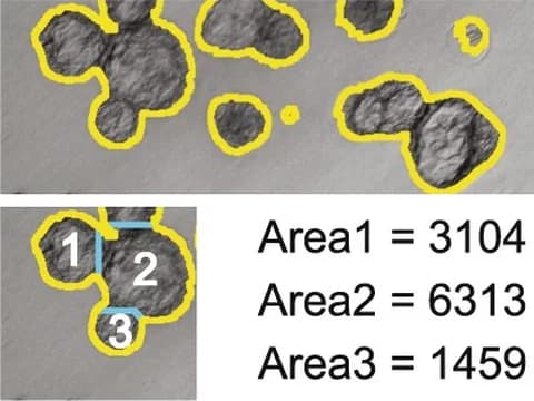 The projected area of each organoid, with its longest and shortest diameters | Every 5 to 15 minutes |
| Growth rate | 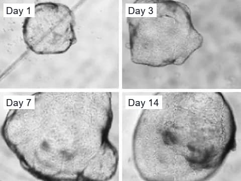 How fast each organoid grows or shrinks, per hour and per day | Every 5 to 15 minutes |
| Shape | 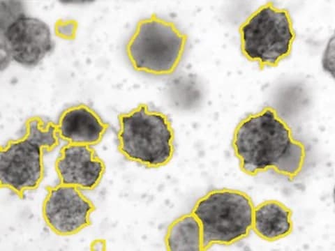 How round, long or irregular each outline is | Every 5 to 15 minutes |
| Movement | 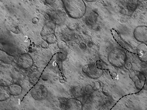 How far and how fast each organoid moves, where organoids are free to move | Every 5 to 15 minutes |
| Fusion | 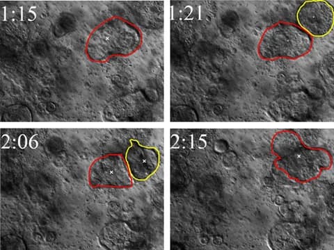 Two organoids joining into one, with both parent IDs kept | Each time it happens |
| Fragmentation | 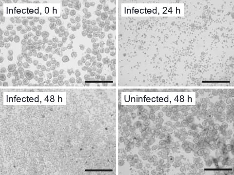 Organoids splitting or shedding pieces, each piece keeping its parent ID | Each time it happens |
| Structural collapse | 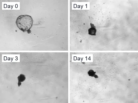 A sudden loss of area, roundness or boundary, with a clip of the change | Each time it happens |
| Brain and neural organoids | ||
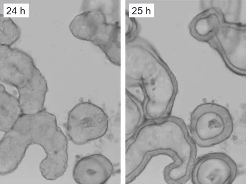
| Outline and folding | 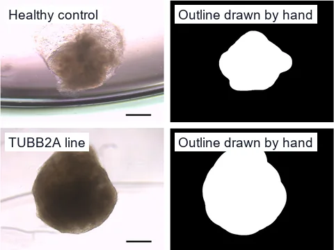 How smooth, lobed or folded each organoid's outline is | Every 5 to 15 minutes |
| Neuroepithelial loops | 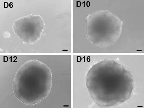 Loops and folds of neuroepithelium along each organoid's edge, where the camera resolves them | Every 5 to 15 minutes |
| Protrusions | 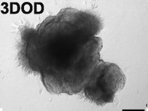 Large protrusions as they form on each organoid's surface, where the camera resolves them | Every 5 to 15 minutes |
| Treatment response | 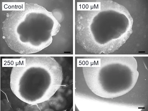 How growth, size and outline change after an exposure, against your controls | Every 5 to 15 minutes |
When a fusion, fragmentation or structural collapse begins, the camera images that well every minute until the change ends. Fragmentation and structural collapse describe what the camera sees in each organoid's outline, not whether its cells are alive.
Beyond the camera
What needs higher magnification or a dye
- Individual neurons and neurites
- Nuclei
- Neural rosettes
- Cortical layers
- Calcium signaling
Evidence
What published studies found in brain organoids
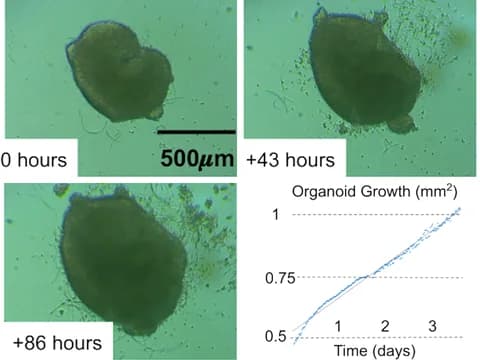
Other research groups measured size and growth changes like these in microscope images of brain organoids.
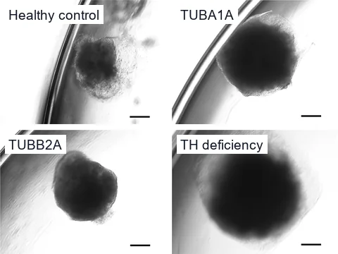
Four lines grew differently
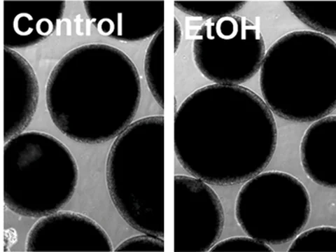
Smaller after ethanol
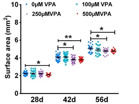
Smaller with valproic acid
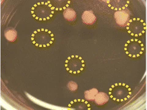
Organoids that failed to develop
Camera specification
The cameras a pilot uses
Around the clock
- A full stack of focal planes for every well, every 5 to 15 minutes
Incubator
- Inside your incubator, or in a chamber beside it that keeps the same conditions
References
- Schröter J, Deininger L, Lupse B, et al. (2024). A large and diverse brain organoid dataset of 1,400 cross-laboratory images of 64 trackable brain organoids. Scientific Data.
- Adams JW, Negraes PD, Truong J, et al. (2023). Impact of alcohol exposure on neural development and network formation in human cortical organoids. Molecular Psychiatry.
- Zang Z, Yin H, Du Z, et al. (2022). Valproic acid exposure decreases neurogenic potential of outer radial glia in human brain organoids. Frontiers in Molecular Neuroscience.
- Gomez-Giro G, Arias-Fuenzalida J, Jarazo J, et al. (2019). Synapse alterations precede neuronal damage and storage pathology in a human cerebral organoid model of CLN3-juvenile neuronal ceroid lipofuscinosis. Acta Neuropathologica Communications.
- Borten MA, Bajikar SS, Sasaki N, et al. (2018). Automated brightfield morphometry of 3D organoid populations by OrganoSeg. Scientific Reports.
- Gulieva RE, Ahmadvand P, Freedman BS (2025). A novel rapalog shows improved safety vs. efficacy in a human organoid model of polycystic kidney disease. Stem Cell Reports.
- Pierzchalska M, Panek M, Grabacka M (2019). The migration and fusion events related to ROCK activity strongly influence the morphology of chicken embryo intestinal organoids. Protoplasma.
- Stroulios G, Brown T, Moreni G, et al. (2022). Apical-out airway organoids as a platform for studying viral infections and screening for antiviral drugs. Scientific Reports.
- Lefferts JW, Kroes S, Smith MB, et al. (2024). OrgaSegment: deep-learning based organoid segmentation to quantify CFTR dependent fluid secretion. Communications Biology.
- Hof L, Moreth T, Koch M, et al. (2021). Long-term live imaging and multiscale analysis identify heterogeneity and core principles of epithelial organoid morphogenesis. BMC Biology.
- Ly VT, Baudin PV, Pansodtee P, et al. (2021). Picroscope: low-cost system for simultaneous longitudinal biological imaging. Communications Biology.
Plan a brain organoid pilot
Tell us about your brain or neural organoids, your plates and the first question you want answered.