ConductVision Organoid Monitor
Organoid models, monitored 24/7
In a pilot, ConductVision records the size, growth, shape and events of every visible organoid around the clock, along with your model's own measures, such as kidney cysts.
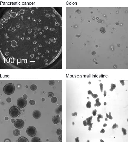
Models
Choose your organoid model
For any other organoid model, a pilot records area, growth, shape, movement and events for every organoid the camera can see, and checks which further measures your images support.
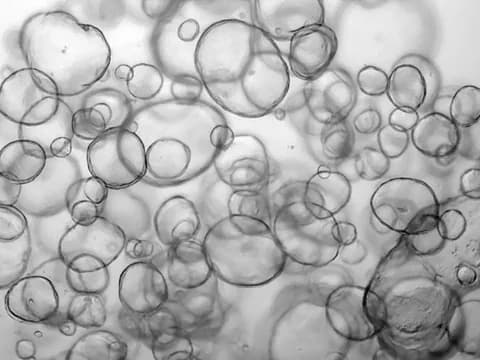
Adapted from Kassis et al. 2019 · CC BY 4.0 Intestinal and colorectal organoids
Growth, buds and lumens for each organoid, including lumens swelling after a treatment.
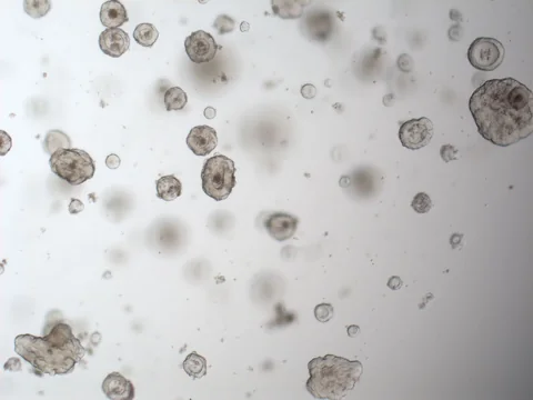
Adapted from Yu et al. 2025 · CC BY 4.0 Tumor organoids
Organoid count per well, growth and regrowth for each organoid, and how responses differ within each well.
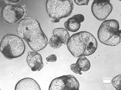
Adapted from Shamshirgaran et al. 2021 · CC BY 4.0 Kidney organoids
Size and shape of each organoid, and its visible cysts as they form, swell and fuse.
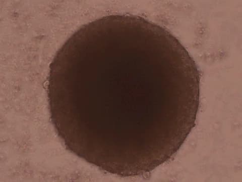
Adapted from Ivanov et al. 2014 · CC BY 4.0 Spheroids and other 3D cultures
Diameter, volume and shape of each spheroid, neurosphere or other cell aggregate, and invasion into a gel.
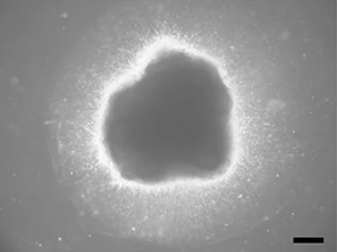
Adapted from Zang et al. 2022 · CC BY 4.0 Brain and neural organoids
Growth, outline and visible folds of each organoid, and how they change after an exposure.
References
- Matthews JM, Schuster B, Kashaf SS, et al. (2022). OrganoID: A versatile deep learning platform for tracking and analysis of single-organoid dynamics. PLOS Computational Biology.
- Kassis T, Hernandez-Gordillo V, Langer R, et al. (2019). OrgaQuant: Human Intestinal Organoid Localization and Quantification Using Deep Convolutional Neural Networks. Scientific Reports.
- Yu H, Lin C, Chen F, et al. (2025). Dose response test of patient-derived cancer organoids to irradiation. Frontiers in Oncology.
- Shamshirgaran Y, Jonebring A, Svensson A, et al. (2021). Rapid target validation in a Cas9-inducible hiPSC derived kidney model. Scientific Reports.
- Ivanov DP, Parker TL, Walker DA, et al. (2014). Multiplexing Spheroid Volume, Resazurin and Acid Phosphatase Viability Assays for High-Throughput Screening of Tumour Spheroids and Stem Cell Neurospheres. PLOS ONE.
- Zang Z, Yin H, Du Z, et al. (2022). Valproic acid exposure decreases neurogenic potential of outer radial glia in human brain organoids. Frontiers in Molecular Neuroscience.
Plan a pilot for your organoid model
Tell us your organoid model, your plates and the first question you want answered.