ConductVision Organoid Monitor
Intestinal and colorectal organoids, monitored 24/7
In a pilot, ConductVision measures the growth, buds and lumens of every visible intestinal or colorectal organoid, from images taken every 5 to 15 minutes.
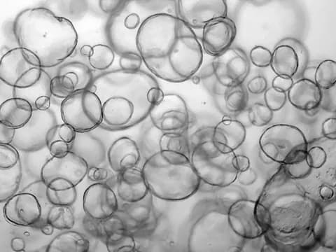
24/7 monitoring
What a pilot records for each intestinal or colorectal organoid
ConductVision records each measure for every visible organoid, keyed by plate, well, organoid ID and time.
| Measure | What it shows | How often |
|---|---|---|
| Every organoid | ||
| Area | 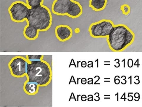 The projected area of each organoid, with its longest and shortest diameters | Every 5 to 15 minutes |
| Growth rate | 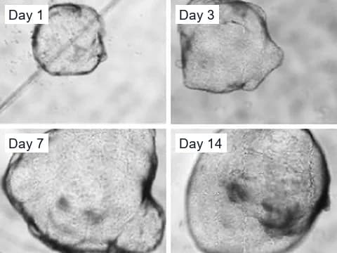 How fast each organoid grows or shrinks, per hour and per day | Every 5 to 15 minutes |
| Shape | 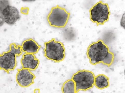 How round, long or irregular each outline is | Every 5 to 15 minutes |
| Movement | 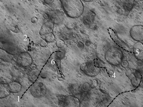 How far and how fast each organoid moves, where organoids are free to move | Every 5 to 15 minutes |
| Fusion | 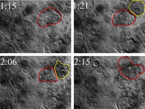 Two organoids joining into one, with both parent IDs kept | Each time it happens |
| Fragmentation | 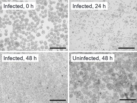 Organoids splitting or shedding pieces, each piece keeping its parent ID | Each time it happens |
| Structural collapse | 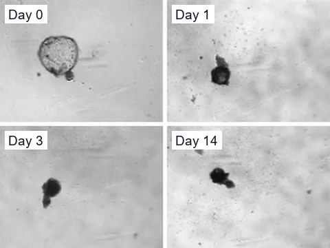 A sudden loss of area, roundness or boundary, with a clip of the change | Each time it happens |
| Intestinal and colorectal organoids | ||
| Buds | 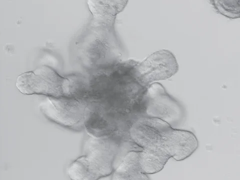 How many gross buds each organoid has, and how fast new ones form | Every 5 to 15 minutes |
| Lumens | 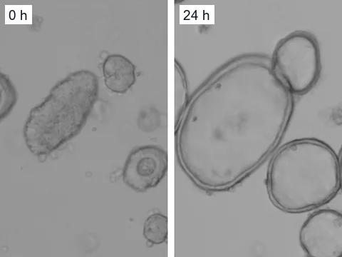 Visible lumens as they form and swell | Every 5 to 15 minutes |
| Cystic or budding | 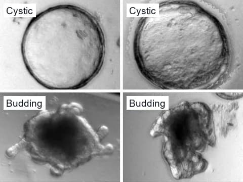 Whether each organoid is a round cyst or a budding organoid | Every 5 to 15 minutes |
| Treatment response | 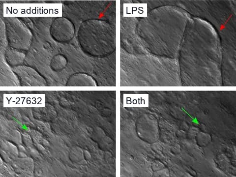 How growth, buds and lumens change after a treatment, against your controls | Every 5 to 15 minutes |
When a fusion, fragmentation or structural collapse begins, the camera images that well every minute until the change ends. Fragmentation and structural collapse describe what the camera sees in each organoid's outline, not whether its cells are alive.
Beyond the camera
What needs higher magnification or a dye
- Individual cells
- Cell polarity
- Fine epithelial layers
- Cell layout inside crypts
Evidence
What published studies found in intestinal organoids
Other research groups measured these in microscope images of intestinal organoids.
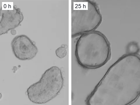
Swelling
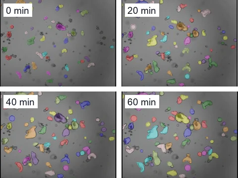
Swelling in brightfield
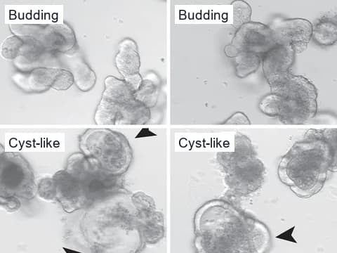
Cystic or budding
SWIFT, a published brightfield pipeline, sorted intestinal organoids into canonical forms, among them cystic, columnar and budding, by area, circularity and brightness.3
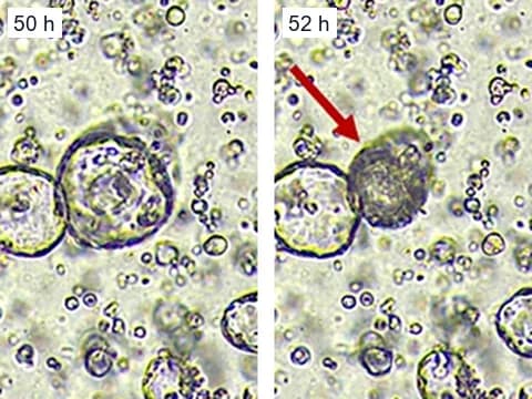
Growth and collapse
Following single organoids over time, the SWIFT authors found that growth rates correlated with Wnt concentration and that rapidly expanding organoids tended to collapse earlier.3
Camera specification
The cameras a pilot uses
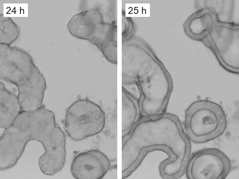
Around the clock
- A full stack of focal planes for every well, every 5 to 15 minutes
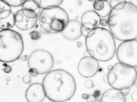
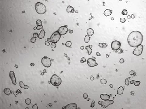
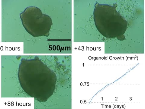
Incubator
- Inside your incubator, or in a chamber beside it that keeps the same conditions
References
- Dekkers JF, Wiegerinck CL, de Jonge HR, et al. (2013). A functional CFTR assay using primary cystic fibrosis intestinal organoids. Nature Medicine.
- Lefferts JW, Kroes S, Smith MB, et al. (2024). OrgaSegment: deep-learning based organoid segmentation to quantify CFTR dependent fluid secretion. Communications Biology.
- Bracq L, Guiet R, Offner S, et al. (2026). A Single-organoid Workflow for quantitative Imaging classiFication and Tracking (SWIFT). Communications Biology.
- Kassis T, Hernandez-Gordillo V, Langer R, et al. (2019). OrgaQuant: Human Intestinal Organoid Localization and Quantification Using Deep Convolutional Neural Networks. Scientific Reports.
- Borten MA, Bajikar SS, Sasaki N, et al. (2018). Automated brightfield morphometry of 3D organoid populations by OrganoSeg. Scientific Reports.
- Gulieva RE, Ahmadvand P, Freedman BS (2025). A novel rapalog shows improved safety vs. efficacy in a human organoid model of polycystic kidney disease. Stem Cell Reports.
- Pierzchalska M, Panek M, Grabacka M (2019). The migration and fusion events related to ROCK activity strongly influence the morphology of chicken embryo intestinal organoids. Protoplasma.
- Stroulios G, Brown T, Moreni G, et al. (2022). Apical-out airway organoids as a platform for studying viral infections and screening for antiviral drugs. Scientific Reports.
- Goveas N, Waskow C, Arndt K, et al. (2021). MLL1 is required for maintenance of intestinal stem cells. PLOS Genetics.
- Yip HYK, Tan CW, Hirokawa Y, et al. (2018). Colon organoid formation and cryptogenesis are stimulated by growth factors secreted from myofibroblasts. PLOS ONE.
- Yan H, Wu T, Li X, et al. (2024). Establishment of the microscope incubation system and its application in evaluating tumor treatment effects through real-time live cellular imaging. Frontiers in Bioengineering and Biotechnology.
- Hof L, Moreth T, Koch M, et al. (2021). Long-term live imaging and multiscale analysis identify heterogeneity and core principles of epithelial organoid morphogenesis. BMC Biology.
- Ly VT, Baudin PV, Pansodtee P, et al. (2021). Picroscope: low-cost system for simultaneous longitudinal biological imaging. Communications Biology.
Plan an intestinal organoid pilot
Tell us about your intestinal or colorectal organoids, your plates and the first question you want answered.