Histology
Prepare consistent slides before the image analysis begins.
Histology should be the hardware-led child hub: tissue processing, embedding, sectioning, staining, and microscopy readiness with a direct handoff into ConductVision.
HISTO-READY / section quality feeds image quality
Histology workflow
A hardware hub organized by the preparation sequence
The page should guide users through how tissue becomes a slide, then show where computer vision can reduce manual review after images are captured.
Prep
Tissue processing and embedding
Match tissue processor, embedding center, cassette, and paraffin workflow to sample volume.
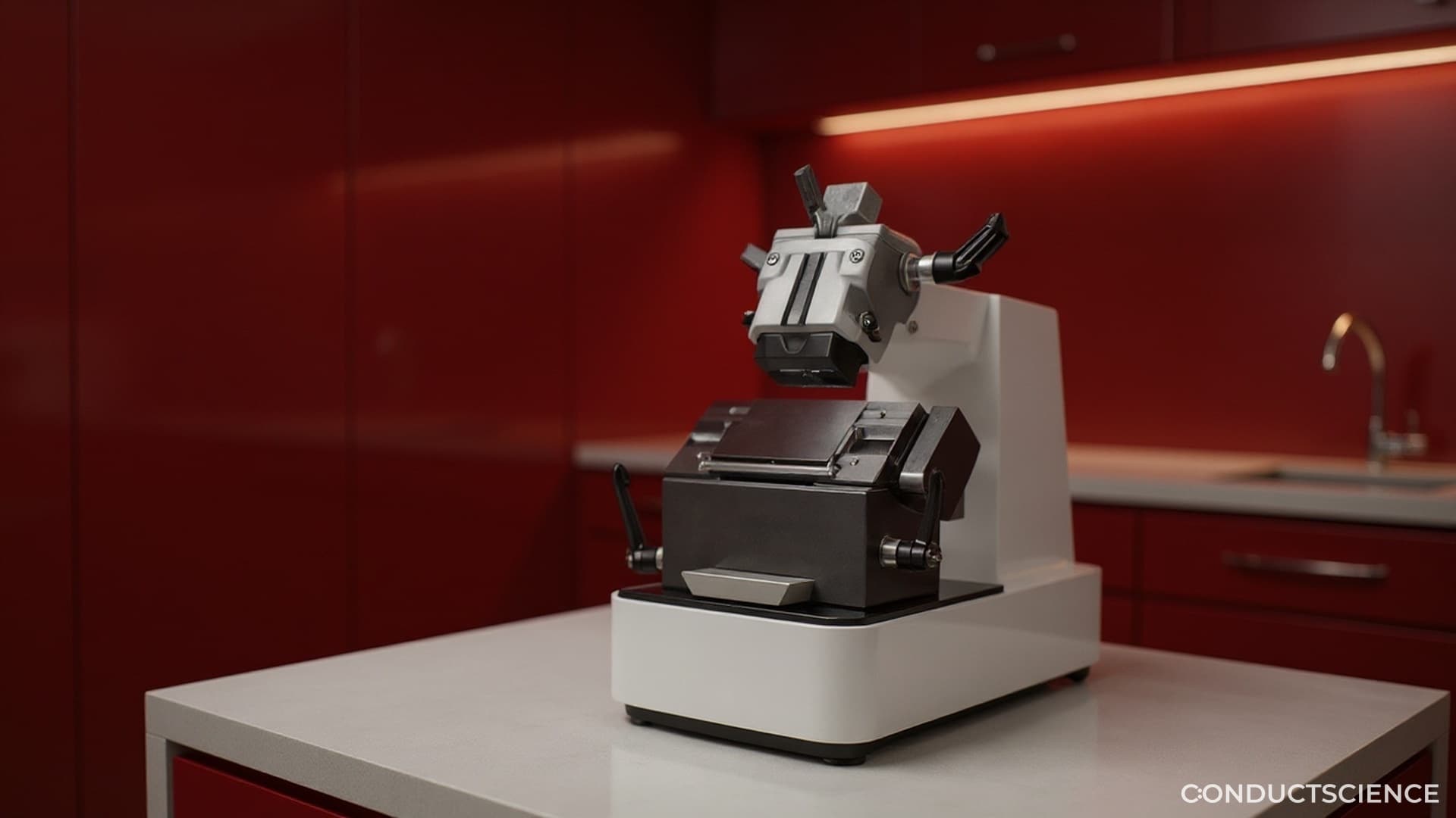
Cut
Sectioning and slide mounting
Select rotary microtomes, cryostats, flotation baths, and drying equipment by throughput.
Stain
Staining and imaging readiness
Standardize slide preparation so H&E, IHC, and fluorescence images are analyzable.
QC
ConductVision handoff
Use image QC and quantification to turn prepared slides into measurable datasets.
Selection logic
Help labs choose the right preparation system
Histology buyers need equipment fit first: sample type, throughput, section thickness, operator load, and imaging endpoint.
Section thickness control
Route users to manual, semi-automatic, automatic, or cryostat systems by sectioning need.
Batch capacity
Connect tissue processor and embedding choices to the number of samples per run.
Slide quality
Explain how processing, sectioning, flotation, and drying affect later CV measurements.
Analysis readiness
Carry users into ConductVision Image when the prepared slide becomes a digital image.
Recommended histology paths
Keep the product buying paths close to the workflow so users can move from education to catalog pages.
Build the slide preparation bench.
Choose the histology instruments first, then connect prepared slides to ConductVision Image for QC and quantification.