Body Composition Imaging Analyzer
Integrated 0.3 T MRI system combining quantitative body composition analysis with imaging and spectroscopy capabilities for non-invasive metabolic phenotyping in laboratory animals.
| Field Strength | 0.3 T (12 MHz) |
| Bore Diameter | 60 mm |
| Modes | Body Composition + MRI + Spectroscopy |
| Automation Level | semi-automated |
| Brand | Greenwaves Scientific |
| Species | Mouse, Rat |
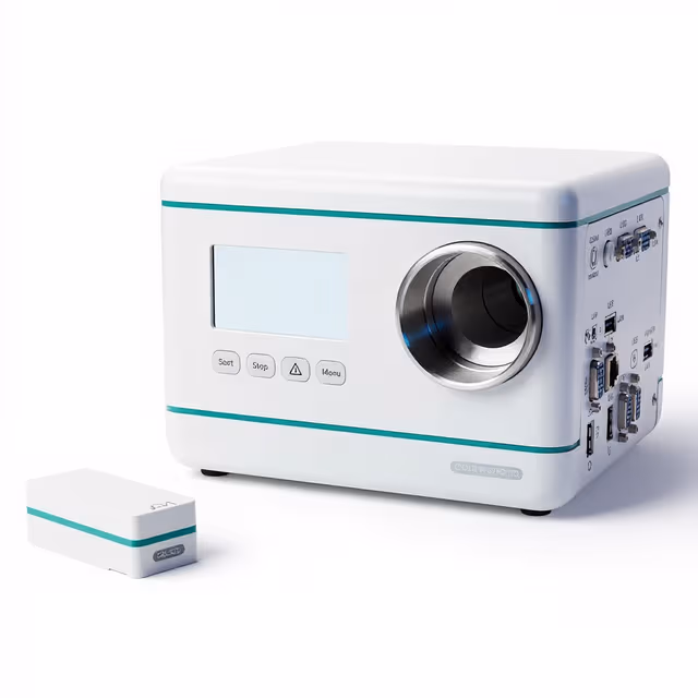
The Body Composition Imaging Analyzer integrates quantitative magnetic resonance body composition measurement with MRI imaging and spectroscopy capabilities in a unified 0.3 T platform. This system enables non-invasive assessment of fat mass, lean mass, and tissue distribution in awake or anesthetized laboratory animals through a 60 mm bore configuration operating at 12 MHz.
The tri-modal functionality allows researchers to perform rapid body composition screening followed by targeted anatomical imaging and spectroscopic analysis of specific tissue regions. This approach supports longitudinal metabolic studies where repeated measurements track adipose tissue changes, organ-specific fat accumulation, and tissue composition alterations over experimental timelines without requiring animal sacrifice or invasive procedures.
How It Works
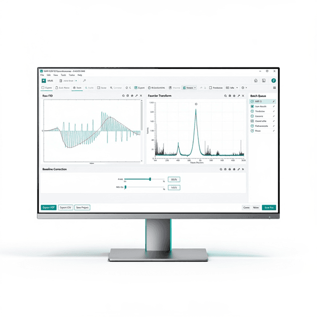
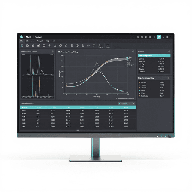
The system operates on nuclear magnetic resonance principles using a 0.3 Tesla permanent magnet to generate radiofrequency pulses at 12 MHz. For body composition analysis, the instrument measures T1 and T2 relaxation times of water and lipid protons throughout the animal's body, with different tissue types exhibiting characteristic relaxation profiles that enable quantitative fat and lean mass determination.
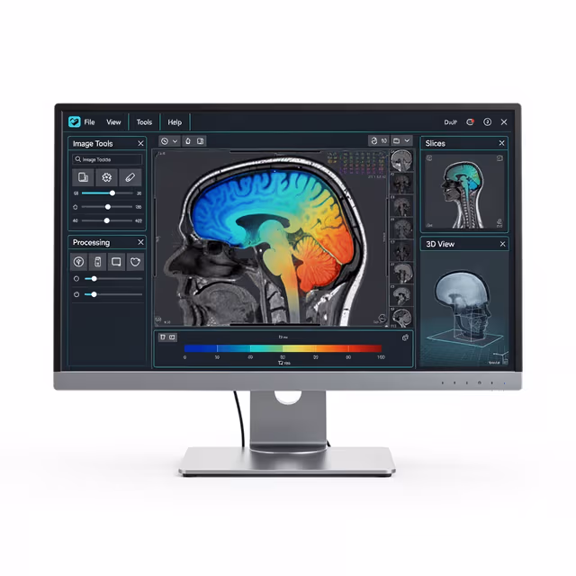
In imaging mode, gradient coils create spatially encoded magnetic field variations that allow anatomical reconstruction through standard MRI pulse sequences. The spectroscopy function analyzes chemical shift differences in the NMR signal to identify specific metabolites and tissue components within defined regions of interest.
The 60 mm bore accommodates animals up to medium rat size while maintaining field homogeneity for accurate quantification. Animals can be measured conscious using restraint tubes or under anesthesia for extended imaging protocols, with the low field strength minimizing heating effects during extended acquisitions.
Features & Benefits
Field Strength
- 0.3 T (12 MHz)
Bore Diameter
- 60 mm
Modes
- Body Composition + MRI + Spectroscopy
Automation Level
- semi-automated
Brand
- Greenwaves Scientific
Research Domain
- Aging Research
- Cardiovascular
- Metabolic Research
- Neurodegeneration
- Pharmaceutical QC
- Toxicology
Species
- Mouse
- Rat
Weight
- 350.0 kg
Dimensions
- L: 100.0 mm
- W: 80.0 mm
- H: 100.0 mm
Comparison Guide
| Feature | This Product | Typical Alternative | Advantage |
|---|---|---|---|
| Field Strength | 0.3 T (12 MHz) | Higher field systems often require anesthesia due to increased acoustic noise and heating effects | Enables awake animal measurements while maintaining sufficient signal quality for body composition analysis. |
| Bore Size | 60 mm diameter | Smaller bore systems may limit animal positioning options | Accommodates standard restraint systems while maintaining field homogeneity for accurate measurements. |
| Operational Modes | Body composition + MRI imaging + spectroscopy | Many systems offer single-mode operation only | Eliminates need for multiple instruments and animal transfers between measurement modalities. |
| Animal State | Awake or anesthetized operation | Higher field systems typically require anesthesia | Supports both high-throughput screening protocols and detailed imaging studies as experimental needs require. |
This system combines quantitative body composition analysis with anatomical imaging and spectroscopy in a 0.3 T platform optimized for laboratory rodent studies. The design enables both awake screening protocols and detailed anesthetized imaging sessions.
Practical Tips
Perform phantom measurements at the beginning of each day before animal studies to verify system stability.
Why: Daily quality assurance detects magnetic field drift that could affect measurement accuracy.
Maintain consistent animal fasting states for body composition studies to reduce measurement variability.
Why: Gastric contents and hydration status can influence tissue quantification results.
Keep the magnet bore clean and free of ferromagnetic debris that could affect field homogeneity.
Why: Metal particles create local field distortions that compromise measurement accuracy.
Remove all ferromagnetic materials from animals and restraint equipment before measurements.
Why: Metal objects pose projectile risks and can cause image artifacts or measurement errors.
Use appropriate signal averaging for your experimental requirements, balancing measurement time with precision needs.
Why: Additional averages improve signal-to-noise ratio but extend acquisition time and animal handling duration.
If measurements appear inconsistent, verify animal positioning reproducibility and check for movement artifacts.
Why: Animal motion during acquisition creates measurement errors that may not be immediately apparent in the data.
Setup Guide
What’s in the Box
- Body Composition Imaging Analyzer main unit
- Animal restraint tubes (multiple sizes) (typical)
- RF coils and positioning accessories (typical)
- Calibration phantoms (typical)
- Acquisition and analysis software (typical)
- Power cables and interface cables (typical)
- User manual and protocol guides (typical)
- Installation and safety documentation (typical)
Warranty
ConductScience provides a comprehensive one-year manufacturer warranty covering parts and technical support. Extended service contracts and calibration services are available for long-term operational support.
Compliance
What is the measurement precision for body composition analysis?
Consult product datasheet for specific precision and accuracy specifications. Precision depends on animal size, measurement protocol, and tissue composition.
Can the system accommodate both mice and rats?
Yes, the 60 mm bore diameter accommodates standard laboratory rodents with appropriate restraint systems for each species.
How long does a typical body composition measurement take?
Consult product datasheet for acquisition times. Duration varies with measurement protocol, signal averaging requirements, and whether imaging is included.
What file formats are provided for data export?
Consult product datasheet for supported data formats and analysis software compatibility requirements.
Is daily calibration required?
Quality assurance measurements with calibration phantoms are recommended for maintaining measurement accuracy and detecting system drift.
Can the system perform spectroscopy on specific tissue regions?
Yes, the tri-modal design enables targeted spectroscopic analysis of user-defined regions of interest following anatomical localization.
What are the space and power requirements?
Consult installation specifications for facility requirements including floor loading, ventilation, and electrical specifications.