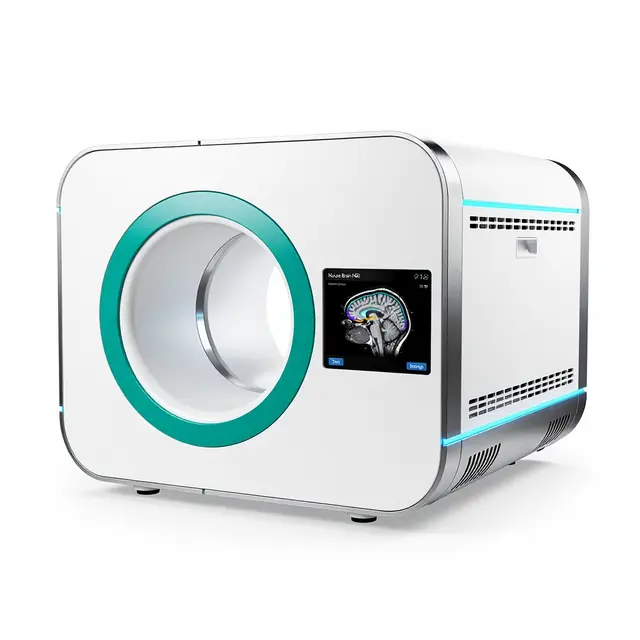
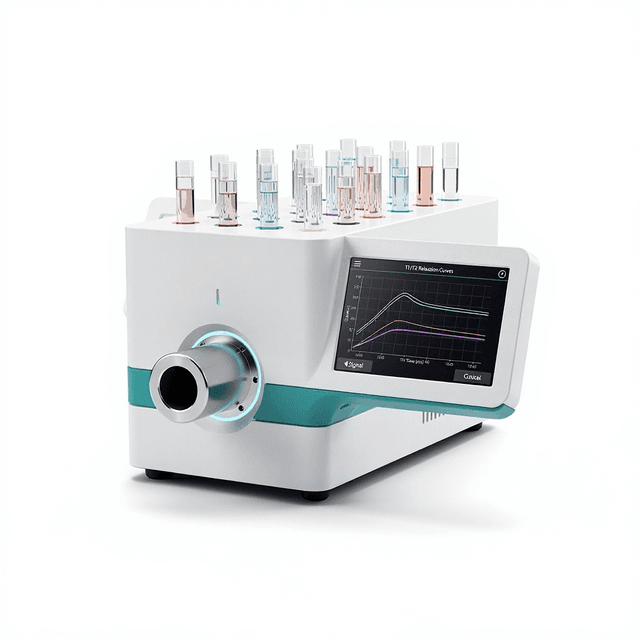
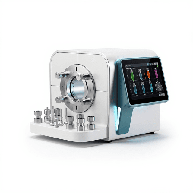
Large-Bore Small Animal MRI System
1.0 T permanent magnet MRI system with 60 mm bore for high-resolution preclinical imaging of rats and small rabbits, supporting comprehensive pulse sequences with up to 100 μm resolution.

Louise Corscadden, PhD
Director of Science · ConductScience
Ask Louise about Large-Bore Small Animal MRI System fit, setup, configuration, or quote prep.
Already working with us? Sign in to connect this with My Scientist.
Key Specifications
Full details →- Model fit
- Rat, Small rabbit
- SKU family
- NMS-NM42
- Sizing
- 4.11 x 2.36 x 1.77 cm
- Ordering
- Online checkout and quote request available
- Category
- MRI Systems
- Build notes
- Confirm accessories, station layout, and support needs before purchase
This large-bore small animal MRI system delivers high-resolution magnetic resonance imaging for preclinical research applications. The system features a 1.0 T permanent magnet with 60 mm bore diameter, providing non-invasive imaging capabilities for rats and small rabbits without the operational costs associated with superconducting systems.
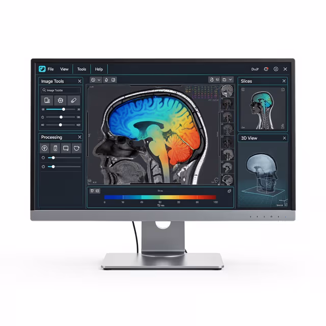
The system operates at 42 MHz frequency and supports comprehensive pulse sequence options including spin echo (SE), multi-slice spin echo (MSE), gradient echo (GRE), inversion recovery (IR), CPMG, spiral imaging (SPI), and additional sequences. With in-plane resolution capabilities up to 100 μm, the system enables detailed anatomical and functional imaging studies for longitudinal research protocols.
How It Works
The system utilizes a permanent magnet design generating a 1.0 Tesla static magnetic field to align nuclear spins in tissue water molecules. Radiofrequency pulses at 42 MHz excite hydrogen nuclei, while gradient coils provide spatial encoding through controlled magnetic field variations. The relaxation of excited nuclei generates measurable RF signals that are digitized and processed through Fourier transformation to reconstruct cross-sectional images.
Multiple pulse sequence options enable tissue contrast optimization through manipulation of repetition time (TR), echo time (TE), and flip angle parameters. Spin echo sequences provide T2-weighted contrast, while gradient echo sequences offer rapid imaging with T1 or T2* weighting. Specialized sequences including inversion recovery enable T1 quantification, and CPMG sequences provide multi-echo T2 analysis for tissue characterization.
The 60 mm bore accommodates animals positioned within dedicated RF coils that optimize signal detection and spatial resolution. Image reconstruction algorithms process the acquired k-space data to generate high-resolution anatomical images with spatial resolution approaching 100 μm in-plane, enabling detailed visualization of tissue microstructure.
Features & Benefits
Field Strength
- 1.0 T (42 MHz)
Bore Diameter
- 60 mm
Animal Size
- Rat
- Small rabbit
Automation Level
- semi-automated
Brand
- Greenwaves Scientific
Research Domain
- Cancer Research
- Cardiovascular
- Developmental Biology
- Neuroscience
- Pharmaceutical QC
- Toxicology
Species
- Rat
Weight
- 0.13 kg
Dimensions
- L: 4.11 mm
- W: 2.36 mm
- H: 1.77 mm
| Feature | This Product | Typical Alternative | Advantage |
|---|---|---|---|
| Magnetic Field Strength | 1.0 T permanent magnet with 42 MHz frequency | Entry-level systems often use 0.5 T fields while high-end research systems operate at 3-9 T | Provides optimal balance of image quality and operational simplicity without cryogenic maintenance requirements. |
| Bore Diameter | 60 mm bore accommodating rats and small rabbits | Compact systems may offer 40-50 mm bores while larger research systems provide 89-120 mm openings | Enables comfortable animal positioning with monitoring equipment while maintaining high spatial resolution through optimized coil design. |
| Spatial Resolution | Up to 100 μm in-plane resolution capability | Basic systems typically achieve 200-300 μm while specialized high-field systems can reach 50-75 μm | Supports detailed morphometric analysis and tissue characterization studies requiring high spatial detail. |
| Pulse Sequence Options | SE, MSE, GRE, IR, CPMG, SPI, and additional sequences | Entry-level systems may offer limited sequence libraries with basic SE and GRE options | Comprehensive sequence availability enables diverse contrast mechanisms and quantitative imaging protocols for varied research applications. |
| Magnet Technology | Permanent magnet design with no cryogen requirements | Research-grade systems commonly use superconducting magnets requiring helium cooling | Eliminates ongoing cryogen costs and provides stable operation without risk of magnet quench events or cooling system failures. |
This system combines practical 1.0 T field strength with comprehensive pulse sequence capabilities and cost-effective permanent magnet operation. The 60 mm bore design optimizes animal accommodation while maintaining high spatial resolution, making it suitable for detailed preclinical imaging studies without superconducting magnet operational complexity.
Practical Tips
Perform magnetic field shimming optimization weekly to maintain image quality and spatial uniformity across the imaging volume.
Why: Permanent magnet systems can experience minor field drift over time affecting image homogeneity and resolution performance.
Monitor RF coil connections and cable integrity regularly to prevent signal degradation and ensure consistent image quality.
Why: Small animal coils experience frequent handling during positioning procedures which can stress connection points.
Use dedicated animal positioning protocols with consistent anesthesia depth monitoring to minimize motion artifacts during acquisition.
Why: Respiratory and cardiac motion can significantly degrade image quality at high spatial resolution settings.
Acquire phantom reference scans at the beginning of each imaging session to verify system performance and image uniformity.
Why: Reference measurements enable detection of system drift and provide baseline data for longitudinal study consistency.
Establish clear magnetic field safety zones around the permanent magnet and screen all personnel and equipment before entering the scan room.
Why: Permanent magnets maintain constant field strength and can attract ferromagnetic objects without warning signs of field activation.
If image artifacts appear, check for ferromagnetic contamination in the imaging area before adjusting sequence parameters.
Why: Metal objects in the magnetic field create characteristic susceptibility artifacts that cannot be corrected through sequence optimization.
Optimize sequence parameters empirically for each contrast agent protocol as relaxivity values vary with field strength.
Why: Contrast agent effectiveness differs significantly between 1.0 T and higher field strengths commonly referenced in literature protocols.
Setup Guide
What’s in the Box
- MRI system console with computer workstation
- Permanent magnet assembly with gradient coils
- RF transmit/receive coil set
- Animal positioning platform (typical)
- Anesthesia delivery accessories (typical)
- Calibration phantom (typical)
- System software and reconstruction packages
- Installation and operation manuals
- Safety documentation and magnetic field maps (typical)
Warranty
ConductScience provides comprehensive warranty coverage including one year parts and labor protection with technical support for system operation and maintenance. Extended service contracts are available for ongoing calibration verification and software updates.
Compliance
What pulse sequences are available for tissue contrast optimization?
The system supports SE, MSE, GRE, IR, CPMG, SPI, and additional sequences enabling T1-weighted, T2-weighted, and T2*-weighted imaging with quantitative parameter mapping capabilities.
How does the permanent magnet design compare to superconducting systems for operational costs?
Permanent magnet systems eliminate ongoing cryogen costs and reduce maintenance requirements compared to superconducting magnets, though field strength is typically lower at 1.0 T versus 3-9 T for superconducting research systems.
What spatial resolution can be achieved for small animal neuroimaging?
The system achieves up to 100 μm in-plane resolution, suitable for detailed anatomical imaging of rat brain structures and morphometric analysis of tissue regions.
Is the bore size adequate for longitudinal studies with animal monitoring?
The 60 mm bore provides sufficient space for rat positioning with anesthesia delivery, physiological monitoring leads, and respiratory gating equipment typically required for longitudinal imaging protocols.
What contrast agents are compatible with this field strength?
Standard gadolinium-based and superparamagnetic iron oxide contrast agents are compatible with 1.0 T imaging, though relaxivity values differ from higher field strengths and should be optimized experimentally.
How does image quality compare between permanent magnet and superconducting systems?
While signal-to-noise ratio is lower at 1.0 T compared to higher field systems, the permanent magnet design provides excellent field homogeneity and stability for consistent image quality in longitudinal studies.
What data formats are supported for image analysis software integration?
Consult product datasheet for specific file format compatibility with common analysis platforms such as ImageJ, MATLAB, and specialized preclinical imaging software packages.
Can functional MRI protocols be implemented on this system?
The gradient echo sequences and temporal resolution capabilities support basic fMRI applications, though sensitivity is reduced compared to higher field strength systems typically used for functional imaging studies.
Have a question about this product?
Have a question? Just ask.
Send it over and we'll email you a personalized answer — no call, no scheduling.
Prefer to talk it through?