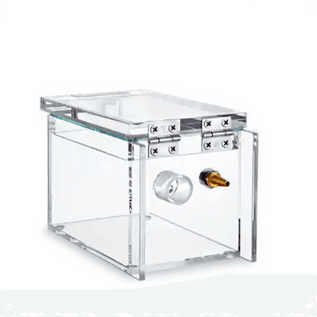
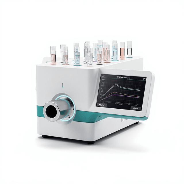
Small Animal MRI System
High-resolution permanent magnet MRI system for preclinical research, available in 1.0T (40H) and 0.5T (60H) configurations with multiple probe options for mice ranging from 1-150g.

Louise Corscadden, PhD
Director of Science · ConductScience
Ask Louise about Small Animal MRI System fit, setup, configuration, or quote prep.
Already working with us? Sign in to connect this with My Scientist.
Key Specifications
Full details →- Model fit
- Mouse
- SKU family
- CS-NM21-060H-I/CS-NM21-040H-I
- Sizing
- 100.0 x 80.0 x 100.0 cm
- Ordering
- Online checkout and quote request available
- Category
- MRI Systems
- Build notes
- Confirm accessories, station layout, and support needs before purchase
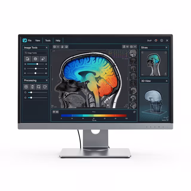
The Small Animal MRI System provides high-resolution magnetic resonance imaging capabilities specifically designed for preclinical research applications. Available in two configurations (Model 40H with 1.0T field strength and Model 60H with 0.5T field strength), the system utilizes permanent magnet technology with precise temperature control and high-performance gradient systems for consistent imaging performance.
The system features dual nonlinear temperature control maintaining magnet stability within ±0.1°C accuracy across a 25-35°C range, with magnetic field stability ≤300Hz/hour. RF capabilities span 1-30MHz with 0.1Hz frequency control accuracy and >300W power amplification. Multiple probe configurations accommodate subjects ranging from 1-150g mice, with effective detection diameters from Ø32mm to Ø62mm depending on probe selection.
Image quality specifications include signal-to-noise ratio ≥20dB, image distortion ≤5%, and image uniformity ≥70%. The system operates on an 8-core CPU industrial control platform with 8GB DDR memory and 1TB storage capacity, providing comprehensive data acquisition and processing capabilities for longitudinal studies and multi-parametric imaging protocols.
How It Works
The Small Animal MRI System operates on the principle of nuclear magnetic resonance, utilizing permanent magnets to generate strong, stable magnetic fields that align hydrogen nuclei in biological tissues. When radiofrequency pulses are applied at specific frequencies (1-30MHz range), these aligned nuclei absorb energy and subsequently emit detectable signals as they return to equilibrium states.
Gradient coils generating >7.5 Gauss/cm (Model 40H) or >5 Gauss/cm (Model 60H) in x, y, and z directions provide spatial encoding by creating controlled magnetic field variations across the imaging volume. The system's dual nonlinear temperature control maintains magnet stability by regulating temperature within ±0.1°C, ensuring consistent field strength and homogeneity over extended imaging sessions.
Signal detection occurs through specialized RF coils matched to different animal sizes, with the system's >300W power amplifier and 2000KHz bandwidth enabling rapid pulse sequences and high-resolution image acquisition. The integrated 8-core processing system handles real-time data collection with maximum CPMG sequences up to 18,000 and minimum echo times of 200μs, supporting diverse imaging protocols from anatomical to functional studies.
Features & Benefits
magnet_field_strength_40H
- 1.0 ± 0.05T
magnet_field_strength_60H
- 0.5±0.05T
magnet_uniformity_40H
- 20 ppm (40mm×40mm×40 mm)
magnet_uniformity_60H
- ≤20ppm (60mm×60mm×60mm)
magnetic_field_stability
- ≤300Hz/Hour
rf_pulse_frequency
- 1-30MHz
frequency_control_accuracy
- 0.1Hz
rf_power_amplifier
- > 300W
maximum_bandwidth
- 2000KHz
mri_gradient_40H
- > 7.5 Gauss/cm for x, y, z
mri_gradient_60H
- > 5 Gauss/cm for x, y, z
probe_sizes_40H
- 40 mm
probe_sizes_60H
- 60 mm, 40 mm or 25 mm (optional)
effective_detection_size_40H
- up to Ø 40 mm
effective_detection_size_60H_60mm
- up to Ø 62mm (45 – 150 g mice)
effective_detection_size_60H_40mm
- up to Ø 42mm (20 – 45 g mice)
effective_detection_size_60H_25mm
- up to Ø 32mm (1 – 20 g mice)
image_quality
- Graphic SNR >= 20db; Image distortion ≤ 5% - Image uniformity >=70%
maximum_data_record_length
- Maximum CPMG is 18,000
minimum_echo_time
- 200 US
computer_specs
- PCI bus industrial control computer platform, 8-core CPU, 8G DDR memory, 1T hard disk
power_frequency
- 50Hz
humidity
- 30-70%
Model
- Model 40H
- Model 60H
Automation Level
- semi-automated
Power/Voltage
- 220V
Temperature Range
- 22-28°C (operating), 25-35°C (magnet temperature control)
Accuracy
- ±0.1°C (temperature accuracy)
Species
- Mouse
Brand
- Greenwaves Scientific
Research Domain
- Cancer Research
- Cardiovascular
- Developmental Biology
- Metabolic Research
- Neuroscience
- Pharmaceutical QC
Weight
- 800.0 kg
Dimensions
- L: 100.0 mm
- W: 80.0 mm
- H: 100.0 mm
| Feature | This Product | Typical Alternative | Advantage |
|---|---|---|---|
| Magnet Technology | Permanent magnet with dual temperature control (±0.1°C accuracy) | Many systems use superconducting magnets requiring cryogenic maintenance | Eliminates ongoing cryogen costs and quench risks while maintaining stable field conditions for consistent imaging. |
| Probe Flexibility | Multiple probe options (25mm, 40mm, 60mm) for 1-150g subject range | Fixed bore systems typically accommodate narrower weight ranges | Enables studies across developmental stages and different mouse strains without compromising image quality. |
| Field Stability | ≤300Hz/hour drift with 20ppm uniformity | Entry-level systems often show higher drift rates | Supports extended imaging protocols and longitudinal studies requiring consistent field conditions. |
| Gradient Strength | >7.5 Gauss/cm (40H) or >5 Gauss/cm (60H) in all three axes | Lower-end systems may offer reduced gradient performance | Provides superior spatial encoding for high-resolution anatomical imaging and accurate volumetric measurements. |
| RF Power and Bandwidth | >300W amplifier with 2000KHz bandwidth and 0.1Hz frequency control | Basic systems often have limited power and frequency precision | Enables advanced pulse sequences and rapid imaging protocols for comprehensive preclinical studies. |
This system combines permanent magnet reliability with advanced RF capabilities and flexible probe configurations, providing field strengths suitable for most preclinical applications while eliminating cryogenic maintenance requirements. The dual temperature control and high gradient strength specifications support consistent, high-quality imaging across diverse research protocols.
| Model | SKU | Listed price | Status | Dimensions |
|---|---|---|---|---|
| Model 60H | CS-NM21-060H-I/CS-NM21-040H-I | Quote | Available | 100.0 x 80.0 x 100.0 cm |
| Model 40H | CS-NM21-060H-I/CS-NM21-040H-I | Quote | Available | 100.0 x 80.0 x 100.0 cm |
Practical Tips
Perform daily field stability verification before imaging sessions and recalibrate RF frequency if drift exceeds 0.1Hz tolerance.
Why: Maintaining precise resonance conditions ensures consistent signal acquisition and reproducible quantitative measurements.
Monitor temperature control system performance weekly and clean temperature sensors monthly to maintain ±0.1°C accuracy.
Why: Temperature stability directly affects magnetic field homogeneity and long-term system performance.
Allow 24-48 hour thermal stabilization after any system shutdown and verify gradient linearity before critical studies.
Why: Thermal equilibrium ensures stable magnetic field conditions and optimal image quality for quantitative analyses.
Select probe size based on subject weight ranges (25mm: 1-20g, 40mm: 20-45g, 60mm: 45-150g) for optimal SNR.
Why: Proper probe matching maximizes signal detection efficiency and image quality for each subject size.
Verify SNR ≥20dB, distortion ≤5%, and uniformity ≥70% using standard phantoms before each experimental series.
Why: Regular quality assurance ensures consistent imaging performance and validates quantitative measurement accuracy.
If image artifacts appear, check gradient coil connections and verify RF shield integrity before adjusting acquisition parameters.
Why: Hardware issues often manifest as systematic image artifacts that cannot be resolved through software adjustments alone.
Maintain minimum 1-meter clearance around magnet assembly and screen all personnel and equipment for ferromagnetic materials.
Why: Permanent magnets maintain constant fields that can attract ferromagnetic objects and pose safety risks even when system is powered off.
Setup Guide
What’s in the Box
- MRI system main unit with permanent magnet assembly
- RF gradient coil system
- Standard probe configuration (40mm for Model 40H, 60mm for Model 60H)
- Industrial control computer with pre-installed software
- Temperature control system components
- Power cables and interface connections
- User manual and technical documentation
- Calibration and performance verification protocols (typical)
Warranty
ConductScience provides comprehensive warranty coverage including one-year manufacturer warranty on all system components with dedicated technical support for installation, calibration, and ongoing maintenance requirements.
Compliance
References
Background reading relevant to this product:
What is the difference in imaging capabilities between the 1.0T and 0.5T field strength models?
The Model 40H (1.0T) provides higher SNR and resolution for detailed anatomical imaging with a 40mm probe, while the Model 60H (0.5T) offers multiple probe options (25mm, 40mm, 60mm) accommodating a broader weight range (1-150g) with slightly reduced gradient strength (>5 vs >7.5 Gauss/cm).
How long does the system require for thermal stabilization before imaging?
The dual nonlinear temperature control system requires 24-48 hours to achieve stable operating conditions with field stability ≤300Hz/hour and temperature accuracy within ±0.1°C across the 25-35°C control range.
What anesthesia protocols are compatible with the extended imaging sessions?
The system design accommodates standard rodent anesthesia protocols including isoflurane delivery systems, though specific anesthesia equipment integration should be verified during installation and consult product datasheet for detailed compatibility specifications.
Can the system perform functional MRI studies in addition to anatomical imaging?
The RF capabilities (1-30MHz range with >300W amplification) and advanced pulse sequence options (18,000 CPMG maximum) support various imaging techniques, though specific fMRI protocol validation would require consultation with technical specifications.
What data formats and analysis software are supported?
The 8-core CPU platform with 1TB storage provides comprehensive data processing, though specific file formats and third-party analysis software compatibility details should be confirmed through product datasheet consultation.
How does the permanent magnet design affect maintenance requirements compared to superconducting systems?
Permanent magnet technology eliminates cryogen refilling, helium quench risks, and associated cryogenic maintenance, requiring only routine temperature control system monitoring and RF calibration verification.
What spatial resolution can be achieved with the different probe configurations?
Resolution capabilities depend on probe selection and imaging parameters, with smaller probes (25mm) providing higher resolution for neonatal subjects while larger probes (60mm) accommodate adult mice; consult technical specifications for detailed resolution parameters.
Have a question about this product?
Have a question? Just ask.
Send it over and we'll email you a personalized answer — no call, no scheduling.
Prefer to talk it through?