Electrophoresis Chip (100 um, Type A)
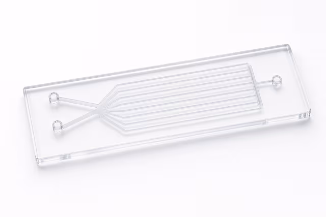
Precision microfluidic chip with 100 × 100 μm channels and cross-injection design for protein separation and electrophoretic analysis.
Reusable chip — designed for multiple experimental runs. Compatible with standard microfluidic tubing: steel pins (0.7 mm ID / 1.0 mm OD) and silicone tubing (0.8 mm ID / 1.9 mm OD). Available in bulk packs (5‑, 10‑, and 25‑unit) for lab-scale and consumable workflows.

Louise Corscadden, PhD
Director of Science · ConductScience
Ask Louise about Electrophoresis Chip (100 um, Type A) fit, setup, configuration, or quote prep.
Already working with us? Sign in to connect this with My Scientist.
Key Specifications
Full details →- Model fit
- 3 selectable configurations
- SKU family
- WHM-0105
- Sizing
- 181.8 x 136.3 x 90.9 cm
- Ordering
- Online checkout and quote request available
- Category
- Electrophoresis Chips
- Build notes
- Confirm accessories, station layout, and support needs before purchase
The Electrophoresis Chip (100 μm, Type A) is a precision microfluidic device engineered for protein separation and electrophoretic analysis in miniaturized format. This cross-injection design chip features 100 × 100 μm channels optimized for high-resolution separation of biomolecules under controlled electric field conditions.
The chip enables researchers to perform capillary electrophoresis separations with reduced sample volumes and enhanced separation efficiency compared to conventional gel-based methods. The cross-injection geometry provides precise sample introduction and separation control, making it suitable for analytical workflows requiring reproducible electrophoretic separation of proteins and other charged biomolecules.
How It Works
Microfluidic electrophoresis separates charged biomolecules based on their size-to-charge ratio under an applied electric field. The chip's cross-injection design creates distinct channels for sample introduction, buffer flow, and separation, enabling precise control over sample plugs and separation conditions.
When voltage is applied across the separation channel, proteins migrate at different velocities based on their electrophoretic mobility. Smaller, more highly charged proteins migrate faster through the 100 μm channels, while larger proteins with lower charge densities migrate more slowly, creating spatial separation. The confined geometry of the microchannels enhances separation efficiency by reducing band broadening and providing consistent electric field distribution.
Detection typically occurs downstream in the separation channel using fluorescence, UV absorbance, or electrochemical methods, allowing real-time monitoring of separated protein bands as they pass the detection zone.
Features & Benefits
Pack Size
- 5-Pack
- 10-Pack
- 25-Pack
Weight
- 3.3 kg
Dimensions
- L: 181.8 mm
- W: 136.3 mm
- H: 90.9 mm
| Feature | This Product | Typical Alternative | Advantage |
|---|---|---|---|
| Channel Cross-Section | 100 × 100 μm square channels | Circular capillaries or larger rectangular channels in basic devices | Square geometry provides more uniform electric field distribution and consistent protein migration behavior. |
| Injection Design | Cross-injection configuration | Simple T-junction or direct injection systems | Enables precise sample plug formation and better separation reproducibility between runs. |
| Application Focus | Optimized for protein separation | General-purpose electrophoresis chips for mixed analytes | Specific design considerations for protein electrophoretic mobility and surface interactions. |
| Standardization | Type A standard configuration | Custom or proprietary chip formats | Ensures compatibility with established microfluidic electrophoresis instrumentation and protocols. |
This chip combines standardized Type A design with protein-specific optimization, offering researchers a reliable platform for miniaturized electrophoretic separations. The cross-injection geometry and 100 μm channels provide precise control over separation conditions while maintaining compatibility with common detection systems.
| Model | SKU | Listed price | Status | Dimensions |
|---|---|---|---|---|
| 25-Pack | WHM-0105-25PK | Quote | Available | 181.8 x 136.3 x 90.9 cm |
| 10-Pack | WHM-0105-10PK | $1,490.00 | Available | 181.8 x 136.3 x 90.9 cm |
| 5-Pack | WHM-0105-5PK | Quote | Available | 181.8 x 136.3 x 90.9 cm |
Practical Tips
Condition new chips with buffer for 30-60 minutes before first use to establish stable electroosmotic flow.
Why: Proper conditioning ensures reproducible migration times and separation efficiency.
Store chips in deionized water or appropriate buffer to prevent channel drying and maintain surface properties.
Why: Dried channels can develop cracks or altered surface chemistry affecting separation performance.
Use standard protein markers to verify separation performance and optimize voltage conditions for your application.
Why: Standardized calibration ensures consistent results and helps identify optimal operating parameters.
Monitor current stability during separations as sudden changes indicate channel problems or bubble formation.
Why: Current fluctuations can indicate channel blockages or air bubbles that compromise separation quality.
If peak broadening occurs, reduce voltage or optimize buffer ionic strength to minimize joule heating effects.
Why: Excessive heating in microchannels can cause thermal gradients that degrade separation resolution.
Always apply voltage with proper electrode placement and avoid touching chip surfaces during operation.
Why: High voltages used in electrophoresis can cause electrical shock and chip contamination affects results.
Setup Guide
What’s in the Box
- Electrophoresis chip (Type A, 100 μm channels)
- Product specification sheet
- Handling and storage guidelines (typical)
Warranty
ConductScience provides standard warranty coverage against manufacturing defects with technical support for proper chip handling and storage procedures.
Compliance
What buffer systems are compatible with this chip design?
Standard electrophoresis buffers including Tris-glycine, bis-tris, and tricine systems work well. Consult product datasheet for specific pH and ionic strength recommendations for optimal protein separation.
How do I prevent channel clogging during protein analysis?
Pre-filter samples through 0.2 μm filters and use appropriate buffer conditioning protocols. Avoid samples with high salt concentrations or particulates that could occlude the 100 μm channels.
What voltage ranges are appropriate for this channel geometry?
Voltage depends on channel length and buffer conductivity. Start with field strengths around 100-300 V/cm and optimize based on separation quality and current stability for your specific application.
Can this chip be reused for multiple separations?
Chips can typically be reused with proper cleaning protocols between runs. Flush thoroughly with cleaning solutions and conditioning buffers to remove protein adsorption and restore surface properties.
What detection methods work with this chip format?
Fluorescence detection is most common, but UV absorbance and electrochemical detection are also compatible. Detection window location depends on your specific chip-to-instrument interface.
How does separation resolution compare to traditional gel electrophoresis?
Microfluidic electrophoresis typically provides higher resolution due to reduced band broadening and more uniform electric fields, though separation capacity may be lower than large-format gels.
Have a question about this product?
Have a question? Just ask.
Send it over and we'll email you a personalized answer — no call, no scheduling.
Prefer to talk it through?