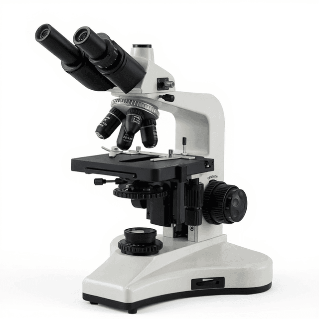
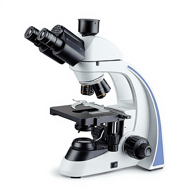
Professional Integrated Coding Nosepiece Upright Trinocular Biological Microscope for Medical Operation & Teaching
Upright trinocular biological microscope with quintuple coding nosepiece, 40X-1000X magnification range, and integrated software recognition for systematic biological specimen examination.
| Automation Level | semi-automated |
| Product Type | Microscope |
| Microscope Type | General Purpose |
| Application | General Laboratory |
| Skill Level | Professional / Research |
| Brand | ConductScience |
The Professional Integrated Coding Nosepiece Upright Trinocular Biological Microscope represents advanced optical instrumentation designed for quantitative biological research and medical education applications. This upright trinocular system combines traditional brightfield microscopy with integrated software-controlled magnification coding, enabling automated objective recognition and systematic documentation of microscopic observations.
The instrument features a quintuple coding nosepiece with RMS international standard threading, supporting magnifications from 40X to 1000X through plan achromatic objectives spanning 4X (NA 0.10) to 100X oil immersion (NA 1.25). The trinocular head configuration accommodates simultaneous visual observation and digital imaging through a 0.5X C-mount interface, while the integrated LED illumination system provides consistent 4500K color temperature illumination equivalent to 100W halogen output.
How It Works
The microscope operates on transmitted light brightfield principles, utilizing a pre-aligned Köhler illumination system with a variable numerical aperture condenser (NA 0.1-1.1). The integrated LED light source provides stable 4500K color temperature illumination, eliminating color temperature drift associated with traditional tungsten-halogen sources. Light passes through the specimen and is collected by plan achromatic objectives with working distances optimized for biological specimens.
The quintuple coding nosepiece incorporates integrated sensors that communicate with PC software to automatically recognize and record objective magnification during specimen examination. This system eliminates manual documentation errors and provides systematic tracking of magnification settings throughout experimental protocols. The trinocular head design splits the optical path with selectable ratios (100:0 or 50:50) between visual observation and camera documentation, enabling real-time observation while maintaining full-resolution digital capture capability.
Features & Benefits
Automation Level
- semi-automated
Product Type
- Microscope
Microscope Type
- General Purpose
Application
- General Laboratory
Skill Level
- Professional / Research
Brand
- ConductScience
Research Domain
- Cell Biology
- Clinical Diagnostics
- Developmental Biology
- Histopathology
- Microbiology
Weight
- 1.9 kg
Dimensions
- L: 4.9 mm
- W: 6.6 mm
- H: 6.6 mm
Comparison Guide
| Feature | This Product | Typical Alternative | Advantage |
|---|---|---|---|
| Nosepiece Capacity | Quintuple (5-position) coding nosepiece with software recognition | Triple or quadruple nosepieces without coding capability | Accommodates complete objective sets with automated magnification tracking for systematic experimental protocols. |
| Illumination System | 10W LED with constant 4500K color temperature | Variable color temperature tungsten-halogen systems | Eliminates color temperature drift and heat generation during extended observation periods. |
| Objective Thread Standard | RMS international standard threading | Proprietary threading systems limit compatibility | Ensures compatibility with standard research objectives from multiple manufacturers for flexible system configuration. |
| Trinocular Design | Adjustable spectral ratios (100:0 or 50:50) with 0.5X C-mount | Fixed ratio trinocular heads or separate camera ports | Provides flexible light distribution for simultaneous visual examination and digital documentation workflows. |
| Condenser Range | Pre-aligned Köhler with NA 0.1-1.1 and scale markings | Fixed aperture or manual alignment condensers | Supports optimal illumination matching across the full magnification range without user alignment procedures. |
This microscope integrates automated objective coding with comprehensive magnification range and trinocular imaging capability. The quintuple nosepiece and RMS threading provide flexibility for diverse research applications, while LED illumination ensures consistent color temperature performance.
Practical Tips
Verify parfocality by focusing at 10X then rotating through all objectives to ensure maintained focus without adjustment.
Why: Proper parfocality calibration prevents specimen damage during magnification changes and maintains consistent focal planes.
Clean objective lenses with appropriate optical tissue and solvents, removing immersion oil completely after each use of the 100X objective.
Why: Oil residue degrades optical performance and can contaminate lower magnification objectives if not properly removed.
Allow LED illumination to stabilize for 10-15 minutes before critical measurements to ensure consistent color temperature.
Why: Initial LED warm-up prevents subtle color variations that could affect quantitative imaging and photomicrography.
Use the coding software to automatically log magnification settings for each image to maintain accurate experimental records.
Why: Automated logging eliminates manual documentation errors and ensures reproducible magnification tracking across experimental sessions.
If objectives fail to code properly, verify clean electrical contacts in the nosepiece and check software connection status.
Why: Dust or contamination on coding contacts can prevent proper objective recognition and disrupt automated documentation workflows.
When using oil immersion, gradually approach the specimen to prevent coverslip breakage due to the minimal 0.21mm working distance.
Why: The short working distance of high-NA objectives requires careful approach to prevent specimen damage and objective contamination.
Adjust interpupillary distance and diopter settings for each user to prevent eye strain during extended observation periods.
Why: Proper ergonomic adjustment reduces user fatigue and maintains observation accuracy during lengthy microscopy sessions.
Store the microscope with dust cover and rotate to the lowest magnification objective to protect higher-power lenses.
Why: Proper storage position protects expensive high-magnification objectives from dust accumulation and accidental damage.
Setup Guide
What’s in the Box
- Trinocular microscope body with integrated LED illumination
- Quintuple coding nosepiece (installed)
- SWH10X23mm eyepieces (pair)
- Plan achromatic objective set (4X, 10X, 20X, 40X, 100X oil)
- Köhler condenser with aperture controls
- Specimen stage with slow rebound clips
- Power supply with regional adapters (typical)
- Microscopy software for objective coding (typical)
- Immersion oil for 100X objective (typical)
- Dust cover and cleaning accessories (typical)
- User manual and alignment instructions (typical)
Warranty
ConductScience provides standard one-year manufacturer warranty covering optical and mechanical components, with technical support for software integration and operational guidance.
Compliance
References
Background reading relevant to this product:
What is the working distance for high-magnification oil immersion objectives?
The 100X oil immersion objective provides 0.21mm working distance with NA 1.25, suitable for standard coverslip specimens with appropriate immersion oil.
How does the coding nosepiece interface with documentation software?
The integrated coding system automatically detects objective position changes and communicates magnification data to PC software, eliminating manual entry errors during systematic observations.
Can the trinocular head accommodate different camera systems?
Yes, the 0.5X C-mount interface accepts standard microscopy cameras, with adjustable spectral ratios allowing simultaneous visual observation and digital capture.
What illumination adjustments are available for different specimen types?
The Köhler condenser provides NA adjustment from 0.1-1.1 with aperture scale markings, supporting brightfield and polarization modes for various specimen contrast requirements.
How is parfocality maintained across the objective range?
Plan achromatic objectives are factory-aligned for parfocality, requiring minimal focus adjustment when rotating between magnifications during specimen examination.
What eyepiece configurations are supported for user comfort?
SWH10X23mm eyepieces provide large field of view with interpupillary adjustment 50-75mm and individual ±5 diopter compensation for extended observation periods.
Can heavy specimens like counting chambers be accommodated?
The slow rebound specimen clips support double slides and heavy samples including cell counting chambers and counting plates without specimen drift during examination.
Have a question about this product?
Accessories
Enhance your setup with compatible accessories

Frequently Bought Together


Related Products