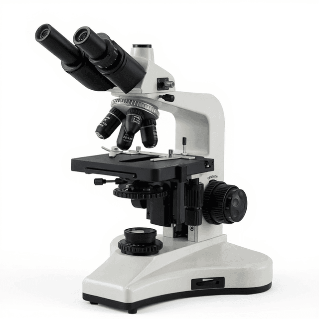
Teaching Microscope
Compound brightfield microscope designed for educational applications and basic research, providing reliable optical performance for specimen observation and morphological analysis.
| Automation Level | manual |
The Teaching Microscope represents a fundamental optical instrument designed for educational and introductory research applications. This compound microscope provides essential magnification capabilities for specimen observation in academic laboratories, teaching environments, and basic research settings. The system employs traditional brightfield illumination with standard objective lenses to deliver clear visualization of prepared samples and biological specimens.
Constructed with educational durability in mind, this microscope offers reliable performance for routine microscopy tasks including cell observation, tissue examination, and basic morphological studies. The compact design (65 x 39 x 29 cm) makes it suitable for shared laboratory spaces while maintaining the optical quality necessary for accurate specimen analysis and documentation.
How It Works
The Teaching Microscope operates on the principle of compound magnification, utilizing a combination of objective and eyepiece lenses to achieve total magnification. Light from the illumination source passes through the condenser system, which focuses illumination onto the specimen mounted on the stage. The objective lens collects light transmitted through or reflected from the specimen, forming a magnified real image at the intermediate image plane.
This intermediate image is further magnified by the eyepiece lens system, producing the final virtual image observed by the user. The total magnification equals the product of the objective lens magnification and eyepiece magnification. The microscope's optical system is designed to provide adequate resolution and contrast for educational applications while maintaining parfocal relationships between objective lenses for efficient specimen examination.
Features & Benefits
Automation Level
- manual
Research Domain
- Cell Biology
- Clinical Diagnostics
- Developmental Biology
- Histopathology
- Microbiology
Weight
- 11.0 kg
Dimensions
- L: 65.0 mm
- W: 39.0 mm
- H: 29.0 mm
Comparison Guide
| Feature | This Product | Typical Alternative | Advantage |
|---|---|---|---|
| Construction Quality | Durable construction designed for educational environments | Entry-level models may use lighter materials with reduced longevity | Maintains alignment and performance under frequent use in teaching laboratories |
| Optical System | Compound brightfield optics with standard magnifications | Basic models may offer fewer objective options or simpler optics | Provides comprehensive magnification range for diverse educational applications |
| Stage Design | Mechanical stage for precise specimen positioning | Simple stages may lack mechanical movement controls | Enables systematic specimen examination and accurate positioning during analysis |
| Footprint Efficiency | Compact 65 x 39 x 29 cm dimensions | Larger systems may require more bench space | Maximizes laboratory space utilization while maintaining full functionality |
This teaching microscope provides reliable compound optics in a durable, compact design suitable for educational environments. The mechanical stage and parfocal objectives support systematic specimen examination workflows common in instructional settings.
Practical Tips
Verify illumination uniformity and objective parfocality at the beginning of each laboratory session.
Why: Ensures consistent image quality and prevents time loss during specimen examination.
Clean objective lenses with lens paper and appropriate solvents, never touching glass surfaces directly.
Why: Optical surface contamination significantly degrades image contrast and resolution.
Start examination with the lowest magnification objective to locate specimens before switching to higher magnifications.
Why: Reduces risk of slide damage and makes specimen navigation more efficient.
If image appears dark or uneven, check condenser alignment and lamp centering before adjusting other parameters.
Why: Illumination problems are the most common cause of poor image quality in brightfield microscopy.
Use consistent illumination settings when comparing specimens or documenting observations.
Why: Standardized conditions improve reproducibility and accuracy of morphological assessments.
Always use appropriate slide thickness and avoid excessive pressure when focusing to prevent objective damage.
Why: High-magnification objectives have very short working distances and can be easily damaged by contact with slides.
Setup Guide
What’s in the Box
- Teaching microscope main unit
- Standard eyepieces (typical)
- Objective lens set (typical)
- Power cord
- Dust cover (typical)
- User manual
- Basic maintenance kit (typical)
Compliance
Warranty & ConductCare
ConductScience provides a standard one-year manufacturer warranty covering defects in materials and workmanship, with technical support available for setup and operational guidance.
What magnification ranges are available with this microscope system?
Consult product datasheet for specific magnification specifications. Total magnification depends on the combination of objective and eyepiece lenses included with the system.
Is this microscope suitable for fluorescence imaging applications?
This is a brightfield microscope designed for transmitted light applications. Fluorescence capability would require specific filter sets and illumination not indicated in the standard configuration.
What type of specimens can be examined with this system?
The microscope is designed for prepared slides including stained tissue sections, blood smears, bacterial cultures, and other transparent or semi-transparent biological specimens suitable for transmitted light examination.
How does image quality compare to research-grade microscopes?
This teaching microscope provides adequate optical quality for educational applications and basic research tasks, though advanced research applications may require higher numerical aperture objectives and specialized contrast methods.
What maintenance is required for consistent performance?
Regular cleaning of optical surfaces, periodic alignment verification, and lamp replacement as needed. Consult user manual for specific maintenance intervals and procedures.
Can digital imaging be integrated with this microscope?
Digital cameras can typically be adapted to standard microscope eyepiece tubes, though specific compatibility depends on camera mount requirements and available adapters.
What is the maximum specimen thickness that can be accommodated?
Standard microscope slides (approximately 1 mm thickness) plus coverslip are typically accommodated. Consult specifications for exact working distance limitations with different objectives.
Have a question about this product?
Accessories
Enhance your setup with compatible accessories


Replacement Parts & Consumables


Frequently Bought Together
Upgrade Options
Related Products