Medical Trinocular Biological Microscope
Research-grade trinocular biological microscope with Kohler illumination, mechanical stage, and high-precision focusing system for detailed specimen examination and documentation.

Louise Corscadden, PhD
Director of Science · ConductScience
Ask Louise about Medical Trinocular Biological Microscope fit, setup, configuration, or quote prep.
Already working with us? Sign in to connect this with My Scientist.
Key Specifications
Full details →- Model fit
- Configured during quote
- SKU family
- BIO-0526
- Sizing
- 49.5 x 35.5 x 26.5 cm
- Ordering
- Online checkout and quote request available
- Category
- Microscopy
- Build notes
- Confirm accessories, station layout, and support needs before purchase
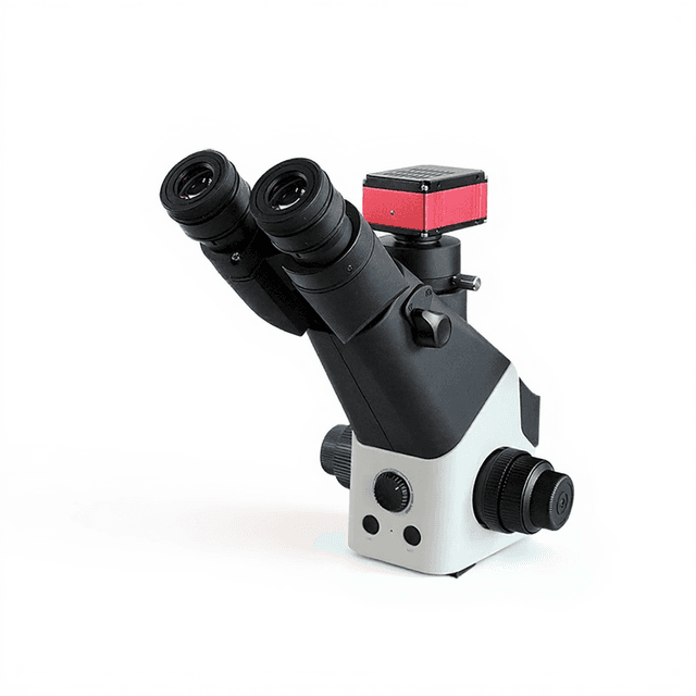
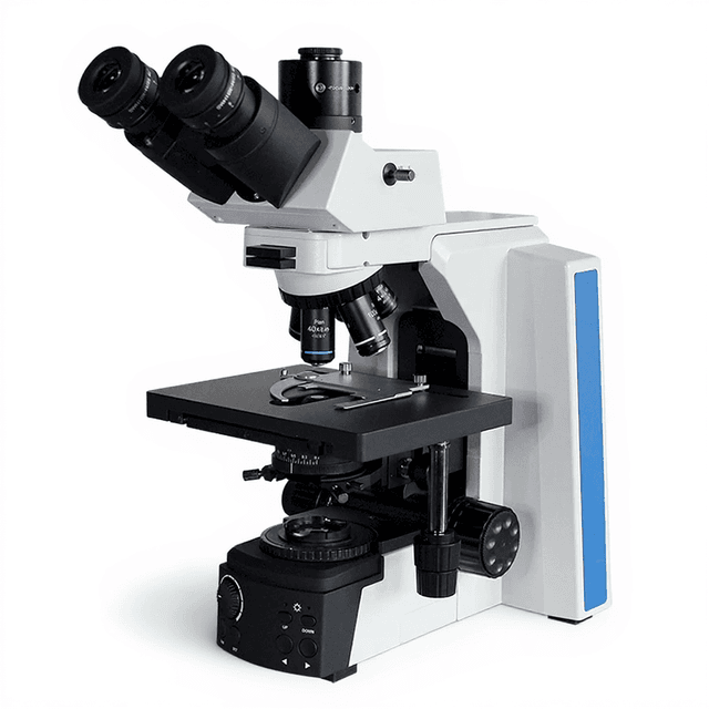
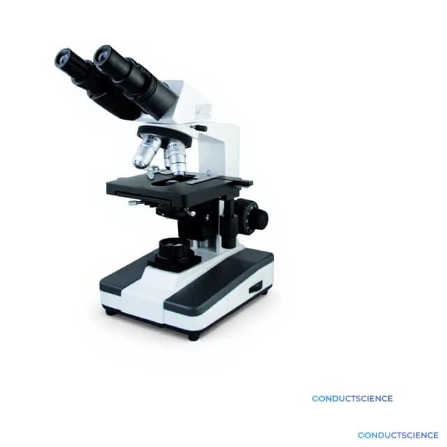
The Medical Trinocular Biological Microscope BIO-0526 is a research-grade brightfield microscope designed for detailed examination of biological specimens across multiple research domains. The instrument features a compensation-free trinocular head with 30° inclination and 360° rotation, enabling comfortable viewing while simultaneously accommodating camera systems for documentation and analysis.
Built around Kohler illumination with halogen lamp (6V/20W) and Abbe condenser (NA 1.25), the microscope delivers consistent, even illumination across the field of view. The coaxial coarse and fine focusing system provides 30mm range with 0.002mm fine division precision, enabling accurate specimen positioning and depth navigation. The mechanical stage (140×140mm) with vernier scale (0.1mm) offers controlled specimen manipulation across a 75×50mm working area, supporting systematic sample examination and precise coordinate recording.
How It Works
The microscope operates on the principle of brightfield illumination, where light from the halogen source passes through the specimen and into the objective lens system. The Kohler illumination system ensures uniform field illumination by focusing the light source image onto the back focal plane of the condenser, while the specimen is illuminated by the front focal plane. This configuration eliminates uneven illumination artifacts and maximizes image contrast.
The Abbe condenser with NA 1.25 concentrates light onto the specimen plane, with the iris diaphragm controlling numerical aperture to optimize resolution and contrast balance. Light transmitted through the specimen is collected by the objective lens and magnified through the eyepiece system, providing total magnifications determined by the objective-eyepiece combination. The trinocular head splits the light path, directing portions to both eyepieces for binocular observation and to the third port for camera attachment.
The coaxial focusing mechanism moves the entire optical assembly vertically relative to the specimen, maintaining parfocal relationships between objectives. Fine focusing adjustments of 0.002mm enable precise optical sectioning through thick specimens, while the mechanical stage coordinate system allows systematic specimen navigation and specific field relocations.
Features & Benefits
Automation Level
- manual
View Head
- Compensation free trinocular head, Inclined at 30°, 360° rotatable, Interpupillary 55~75mm
Eyepiece
- P10X/18mm, P16X/11mm
Focusing
- Coaxial Coarse&Fine Adjustable System, Range 30mm, Fine Division 0.002mm
Stage
- Mechanical Stage 140*140mm, Moving Range 75*50mm, Vernier 0.1mm
Condenser
- Abbe NA1.25 condenser with Iris Diaphragm&Filter
Illumination
- Halogen lamp 6V/20W, Kohler illumination, brightness adjustable
Filter
- Blue, yellow, green
Package Size
- 355*265*495mm
Brand
- ConductScience
Research Domain
- Analytical Chemistry
- Cell Biology
- Clinical Diagnostics
- Developmental Biology
- Histopathology
- Microbiology
Power/Voltage
- AC85~240V, 50/60Hz
Weight
- 7.5kg
Weight
- 7.5 kg
Dimensions
- L: 49.5 mm
- W: 35.5 mm
- H: 26.5 mm
| Feature | This Product | Typical Alternative | Advantage |
|---|---|---|---|
| Head Configuration | Compensation-free trinocular head with 30° inclination and 360° rotation | Entry-level models typically offer binocular heads without camera ports | Enables simultaneous visual observation and digital documentation without optical compromises. |
| Focusing Precision | Coaxial focusing system with 0.002mm fine division | Basic models often provide 0.01mm or coarser focusing increments | Provides precise optical sectioning control essential for high-magnification cellular analysis. |
| Stage System | Mechanical stage 140×140mm with 75×50mm movement range and 0.1mm vernier scale | Lower-cost microscopes frequently use plain stages without mechanical controls | Enables systematic specimen scanning and precise coordinate recording for quantitative work. |
| Condenser Specification | Abbe condenser NA 1.25 with iris diaphragm and filter holder | Entry-level models commonly feature fixed condensers with lower numerical apertures | Maximizes resolution potential and provides aperture control for optimal contrast balance. |
| Illumination System | Kohler illumination with 6V/20W halogen lamp and brightness adjustment | Basic microscopes often use critical illumination with limited intensity control | Delivers uniform field illumination essential for professional documentation and analysis. |
The BIO-0526 combines research-grade optical performance with practical features like trinocular documentation capability and mechanical stage precision. The Kohler illumination system and high-NA condenser deliver professional image quality, while the coaxial focusing system provides the precision needed for detailed cellular analysis across multiple research applications.
Practical Tips
Perform Kohler illumination setup each time you change objectives or begin a new session to ensure optimal image quality.
Why: Proper condenser alignment and aperture settings are critical for maximum resolution and contrast.
Clean objective lenses weekly using lens paper and appropriate cleaning solution, working from center outward in circular motions.
Why: Oil, dust, and specimen residue on objectives significantly degrade image quality and resolution.
Start examination at lowest magnification and work upward, using mechanical stage controls rather than moving slides by hand.
Why: This prevents damage to specimens and objectives while maintaining precise positioning for systematic analysis.
If image appears dim or uneven, check that condenser is properly centered and aperture diaphragm is not overly restricted.
Why: Incorrect condenser alignment or excessive aperture restriction are common causes of poor illumination uniformity.
Use appropriate filters (blue, yellow, green) to enhance contrast for different staining methods and specimen types.
Why: Proper filter selection improves visibility of cellular details and reduces eye strain during extended observation.
Allow halogen lamp to cool for several minutes before handling or replacing bulbs, and avoid touching bulb surfaces with bare hands.
Why: Hot bulbs can cause burns, and oil from skin contact reduces lamp life and can cause premature failure.
Record stage coordinates when documenting specific fields to enable precise field relocation for follow-up observations.
Why: The vernier scale system allows reproducible positioning essential for longitudinal studies and multi-user workflows.
Cover the microscope with dust cover when not in use and store in stable temperature environment away from vibration sources.
Why: Environmental protection preserves optical alignment and prevents contamination of precision mechanical components.
Setup Guide
What’s in the Box
- Medical trinocular biological microscope main unit
- P10X/18mm eyepieces (pair)
- P16X/11mm eyepieces (pair)
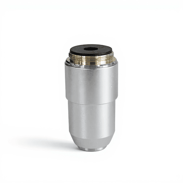
- Objectives set (typical)
- Abbe condenser with iris diaphragm
- Blue, yellow, and green filters
- AC power cord
- Dust cover (typical)
- User manual and documentation (typical)
Warranty
ConductScience provides a standard one-year manufacturer warranty covering defects in materials and workmanship, with technical support available for setup, calibration, and operational guidance throughout the warranty period.
Compliance
References
Background reading relevant to this product:
What magnification combinations are available with the included eyepieces?
The microscope includes P10X/18mm and P16X/11mm eyepieces. Total magnification depends on objective lenses used - with standard 4X, 10X, 40X, and 100X objectives, you achieve magnifications from 40X to 1600X. The 16X eyepieces provide higher magnification for detailed examination while the 10X eyepieces offer wider fields of view.
How do I achieve proper Kohler illumination setup?
Focus the specimen using low magnification, then close the field diaphragm and adjust condenser height until the diaphragm edges are sharp and centered. Open the field diaphragm to just fill the field of view, then adjust the aperture diaphragm to approximately 2/3 of the objective's numerical aperture for optimal contrast.
What is the working distance and stage capacity for large specimens?
The mechanical stage platform is 140×140mm with specimen movement range of 75×50mm. Working distance varies by objective lens used. The stage can accommodate standard slides and larger specimens within the platform dimensions, with vernier scales providing 0.1mm positioning precision.
Can this microscope accommodate oil immersion objectives?
Yes, the Abbe condenser with NA 1.25 and standard objective mount support oil immersion techniques. The condenser's high numerical aperture is specifically designed to work with oil immersion objectives for maximum resolution at high magnifications.
How do I attach a camera to the trinocular head?
The trinocular head includes a third port specifically designed for camera attachment. Consult the product datasheet for specific mounting thread specifications and adapter requirements, as these vary by camera model and sensor type.
What maintenance is required for the halogen illumination system?
The 6V/20W halogen lamp should be replaced when brightness significantly diminishes or color temperature shifts. Handle replacement bulbs with clean cloth to avoid oil contamination, and allow cooling time before handling. Clean condenser and filter surfaces regularly to maintain optimal illumination quality.
How does this compare to fluorescence microscopy systems?
This is a brightfield microscope optimized for transmitted light examination of stained specimens. Unlike fluorescence systems, it cannot excite fluorophores or detect fluorescent emissions. However, it provides superior resolution for morphological analysis and works with all standard histological stains without specialized filter cubes.
Have a question about this product?
Have a question? Just ask.
Send it over and we'll email you a personalized answer — no call, no scheduling.
Prefer to talk it through?
Accessories
Enhance your setup with compatible accessories


Replacement Parts & Consumables


Frequently Bought Together

Related Products