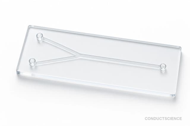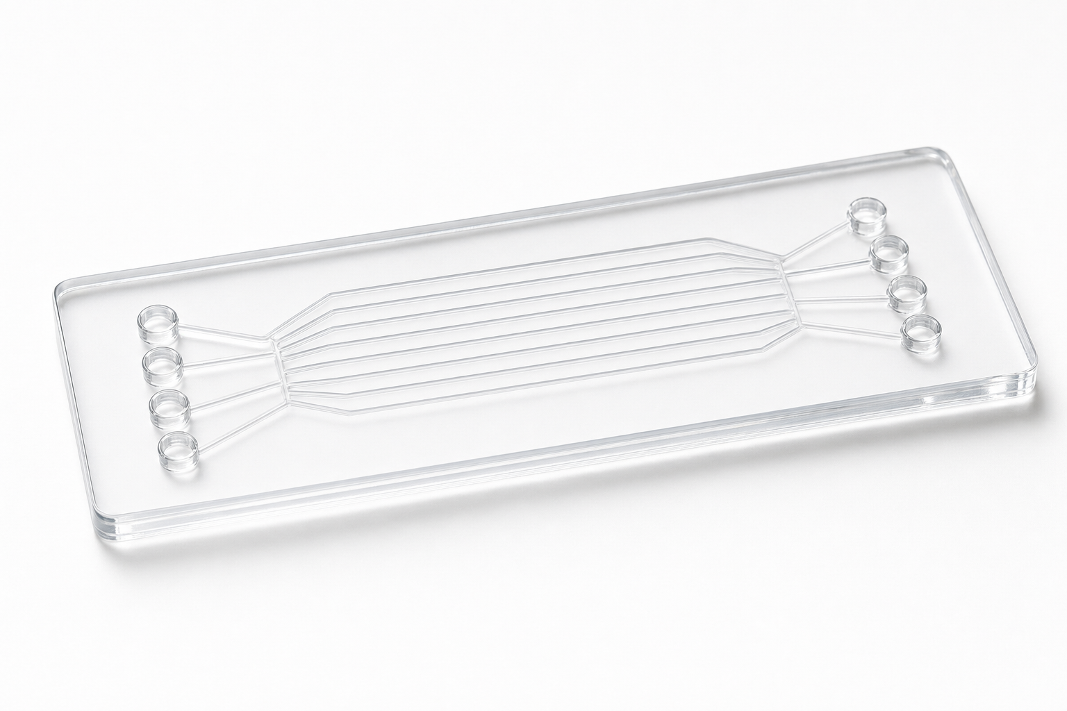
8-Port PDMS Drug Screening Chip (40 um)
Eight-port PDMS microfluidic chip with 40 x 40 μm channels designed for parallel drug screening and dose-response assays with minimal reagent consumption.
Reusable chip — designed for multiple experimental runs. Compatible with standard microfluidic tubing: steel pins (0.7 mm ID / 1.0 mm OD) and silicone tubing (0.8 mm ID / 1.9 mm OD). Available in bulk packs (5‑, 10‑, and 25‑unit) for lab-scale and consumable workflows.

Louise Corscadden, PhD
Director of Science · ConductScience
Ask Louise about 8-Port PDMS Drug Screening Chip (40 um) fit, setup, configuration, or quote prep.
Already working with us? Sign in to connect this with My Scientist.
Key Specifications
Full details →- Model fit
- 3 selectable configurations
- SKU family
- WHM-0084
- Sizing
- 181.8 x 136.3 x 90.9 cm
- Ordering
- Online checkout and quote request available
- Category
- Drug Screening Chips
- Build notes
- Confirm accessories, station layout, and support needs before purchase
The 8-Port PDMS Drug Screening Chip features 40 x 40 μm channels fabricated in polydimethylsiloxane (PDMS), providing a versatile microfluidic platform for pharmaceutical research applications. The chip architecture supports multiple simultaneous assays through eight distinct ports, enabling parallel testing of drug compounds or concentration gradients within a single experimental run.
This microfluidic device is designed for cell-based drug screening assays where precise fluidic control and minimal reagent consumption are critical. The 40 μm channel dimensions accommodate various cell types while maintaining laminar flow characteristics essential for reproducible dose-response studies. The PDMS construction ensures optical transparency for real-time microscopy and provides surface properties suitable for cell adhesion and culture applications.
How It Works
The 8-Port PDMS Drug Screening Chip operates on microfluidic principles where fluid flow is governed by pressure differentials and channel geometry. The 40 x 40 μm square channels maintain laminar flow conditions due to low Reynolds numbers, preventing turbulent mixing and enabling precise spatial control of chemical environments. Each port serves as either an inlet or outlet, allowing researchers to establish multiple independent flow paths or create interconnected channel networks depending on experimental requirements.
Drug compounds introduced through different ports can be diluted and mixed within the channel network to generate concentration gradients. The PDMS material properties facilitate gas exchange while maintaining aqueous solution containment. Cell adhesion occurs on the channel surfaces, where adherent cells are exposed to flowing media containing test compounds. Real-time monitoring is achieved through the optically transparent PDMS, enabling fluorescence or brightfield microscopy to track cellular responses.
The microfluidic format reduces reagent consumption compared to traditional well-plate assays while providing enhanced control over the cellular microenvironment. Shear stress from fluid flow can be adjusted by modifying flow rates, and the small channel dimensions enable rapid equilibration of drug concentrations around cultured cells.
Features & Benefits
Pack Size
- 5-Pack
- 10-Pack
- 25-Pack
Weight
- 3.3 kg
Dimensions
- L: 181.8 mm
- W: 136.3 mm
- H: 90.9 mm
| Feature | This Product | Typical Alternative | Advantage |
|---|---|---|---|
| Number of Ports | 8 ports for parallel testing | Single or dual-port designs common in basic microfluidic chips | Enables higher-throughput screening within a single chip experiment while maintaining independent flow control |
| Channel Dimensions | 40 x 40 μm square channels | Varies by model, often rectangular or circular profiles | Square cross-section provides consistent surface area for cell attachment and predictable flow characteristics |
| Material Construction | PDMS fabrication | Glass or polymer alternatives available | PDMS offers optimal combination of optical transparency, gas permeability, and biocompatibility for cell culture applications |
| Application Focus | Designed specifically for drug screening and dose-response assays | General-purpose microfluidic devices require custom adaptation | Port configuration and channel dimensions optimized for pharmaceutical research workflows without additional customization |
This 8-port PDMS chip provides a specialized solution for drug screening applications with parallel testing capabilities and channel dimensions optimized for cell-based assays. The multi-port architecture enables comprehensive dose-response studies while the PDMS construction ensures compatibility with standard cell culture and microscopy protocols.
| Model | SKU | Listed price | Status | Dimensions |
|---|---|---|---|---|
| 25-Pack | WHM-0084-25PK | Quote | Available | 181.8 x 136.3 x 90.9 cm |
| 10-Pack | WHM-0084-10PK | $1,490.00 | Available | 181.8 x 136.3 x 90.9 cm |
| 5-Pack | WHM-0084-5PK | Quote | Available | 181.8 x 136.3 x 90.9 cm |
Practical Tips
Verify flow rates through each port using fluorescent dyes and time-lapse imaging before cell experiments.
Why: Channel fabrication variations can create flow rate differences between ports that affect experimental reproducibility.
Flush channels with PBS after each use and store in sterile conditions if planning limited reuse.
Why: Proper cleaning prevents biofilm formation and maintains channel patency for subsequent experiments.
Pre-condition PDMS surfaces with serum-containing media for 1-2 hours before cell seeding.
Why: Protein adsorption reduces non-specific binding and improves cell attachment consistency across channels.
If cells detach during perfusion, reduce flow rates and verify surface coating uniformity.
Why: Excessive shear stress or inadequate surface treatment can cause cell loss during drug exposure phases.
Include control channels with media-only perfusion in each experiment for baseline comparisons.
Why: Flow-induced effects on cell behavior must be distinguished from drug-specific responses in data analysis.
Use appropriate containment protocols when testing cytotoxic compounds in the microfluidic format.
Why: Small volumes can concentrate toxic vapors, and PDMS permeability may allow compound migration outside channels.
Monitor temperature stability when using stage-top incubators with perfusion systems.
Why: Temperature fluctuations affect both cell viability and drug stability in long-term screening assays.
Image the same regions consistently across time points to track individual cell responses.
Why: Spatial variations in flow patterns and drug concentrations require location-specific analysis for accurate dose-response curves.
Setup Guide
What’s in the Box
- 8-Port PDMS Drug Screening Chip
- User manual with protocols (typical)
- Quality control certificate (typical)
Warranty
ConductScience provides a standard manufacturer warranty covering defects in materials and workmanship. Technical support includes protocol guidance and troubleshooting assistance for optimal experimental results.
Compliance
What cell types are compatible with the 40 μm channel height?
The 40 μm channels accommodate most adherent cell lines including HeLa, HEK293, and primary cell cultures. Suspension cells can also be perfused, though adherent protocols provide better spatial control.
How do I generate concentration gradients across the eight ports?
Connect alternating ports to different drug concentrations and utilize diffusion mixing within channel networks, or use external mixing devices to create gradient inputs at specific ports.
What flow rates are recommended for drug screening assays?
Typical flow rates range from 0.1-10 μL/min depending on cell type and assay duration. Lower rates (0.1-1 μL/min) suit long-term culture, while higher rates enable rapid compound exchange.
Can the chip be reused for multiple experiments?
The PDMS chip is designed for single-use applications to prevent cross-contamination. Thorough cleaning protocols may enable limited reuse, but performance consistency requires fresh chips per experiment.
What microscopy techniques work with this PDMS format?
The transparent PDMS supports brightfield, phase contrast, fluorescence, and live-cell imaging. Working distances up to 2 mm accommodate most objective lenses for high-resolution imaging.
How long can cells survive in the microfluidic environment?
With appropriate flow rates and media perfusion, cells typically maintain viability for 24-72 hours. The PDMS gas permeability helps maintain physiological oxygen and CO2 levels.
What drug concentrations can be tested simultaneously?
The eight ports enable testing up to eight different concentrations or compounds per experiment. Concentration ranges depend on your stock solutions and dilution scheme design.
Have a question about this product?
Have a question? Just ask.
Send it over and we'll email you a personalized answer — no call, no scheduling.
Prefer to talk it through?