Cell Culture Observation Microfluidic Chip
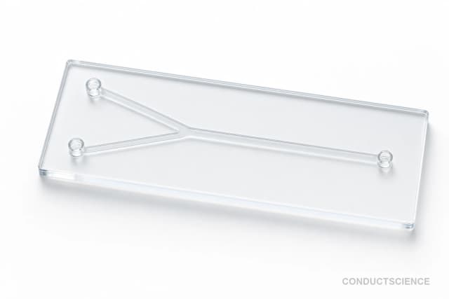
PMMA-based microfluidic chip designed for live cell imaging and observation, enabling real-time monitoring of cellular behavior and dynamics under microscopic analysis.
Reusable chip — designed for multiple experimental runs. Compatible with standard microfluidic tubing: steel pins (0.7 mm ID / 1.0 mm OD) and silicone tubing (0.8 mm ID / 1.9 mm OD). Available in bulk packs (5‑, 10‑, and 25‑unit) for lab-scale and consumable workflows.

Louise Corscadden, PhD
Director of Science · ConductScience
Ask Louise about Cell Culture Observation Microfluidic Chip fit, setup, configuration, or quote prep.
Already working with us? Sign in to connect this with My Scientist.
Key Specifications
Full details →- Model fit
- 3 selectable configurations
- SKU family
- WHM-0075
- Sizing
- 181.8 x 136.3 x 90.9 cm
- Ordering
- Online checkout and quote request available
- Category
- Cell Culture Chips
- Build notes
- Confirm accessories, station layout, and support needs before purchase
The Cell Culture Observation Microfluidic Chip is a PMMA-based microfluidic device designed for live cell imaging and observation applications. This transparent chip provides researchers with a controlled microenvironment for studying cellular behavior, growth patterns, and dynamic processes under microscopic observation. The chip's optical clarity and biocompatible PMMA construction enable real-time monitoring of cell cultures with minimal interference to natural cellular processes.
The device supports various cell observation protocols including cell migration assays, morphology analysis, and time-lapse microscopy studies. Its microfluidic design allows for precise control of the cellular microenvironment while maintaining compatibility with standard microscopy platforms. The chip facilitates detailed analysis of cellular dynamics, growth monitoring, and behavioral studies in a controlled laboratory setting.
How It Works
The microfluidic chip operates on the principle of controlled fluid flow within microscale channels fabricated in PMMA substrate. The transparent polymer construction allows unobstructed optical access for microscopic observation while providing a sterile, controlled environment for cell culture. Cells are introduced into the chip's chambers or channels where they can attach, grow, and be monitored over extended periods.
The microfluidic design enables precise control of nutrient delivery, waste removal, and environmental conditions through regulated fluid flow. The chip's geometry creates predictable flow patterns that can be used to study cell responses to chemical gradients, shear stress, or other environmental stimuli. The PMMA material provides excellent optical clarity across visible wavelengths while maintaining biocompatibility for cell culture applications.
Observation is conducted using standard microscopy techniques including bright field, phase contrast, or fluorescence imaging. The chip's thin profile and flat surfaces minimize optical distortion and enable high-resolution imaging of cellular processes. Time-lapse imaging capabilities allow researchers to track dynamic cellular behaviors over hours or days of continuous observation.
Features & Benefits
Pack Size
- 5-Pack
- 10-Pack
- 25-Pack
Weight
- 3.3 kg
Dimensions
- L: 181.8 mm
- W: 136.3 mm
- H: 90.9 mm
| Feature | This Product | Typical Alternative | Advantage |
|---|---|---|---|
| Substrate Material | PMMA construction | Glass or PDMS substrates are common alternatives | PMMA provides good optical clarity with lower cost and easier handling compared to glass, while offering better rigidity than PDMS |
| Optical Clarity | Transparent PMMA for microscopic observation | Varies by material choice and fabrication quality | Enables high-resolution imaging across multiple microscopy modalities with minimal optical interference |
| Application Focus | Specialized for cell observation and live imaging | Generic microfluidic platforms may lack optimization for imaging applications | Purpose-built design optimizes channel geometry and optical access for cellular observation protocols |
| Biocompatibility | PMMA biocompatible construction | Material biocompatibility varies by substrate choice | Supports extended cell culture periods with minimal cytotoxic effects on cultured cells |
| Sterilization Options | UV and low-temperature sterilization compatible | Sterilization methods depend on substrate thermal properties | Multiple sterilization approaches accommodate different laboratory protocols and equipment availability |
This PMMA-based microfluidic chip provides a specialized platform for cell observation applications with optimized optical properties and biocompatibility. The design focuses specifically on live imaging requirements while maintaining practical advantages in cost and handling compared to alternative substrate materials.
| Model | SKU | Listed price | Status | Dimensions |
|---|---|---|---|---|
| 25-Pack | WHM-0075-25PK | Quote | Available | 181.8 x 136.3 x 90.9 cm |
| 10-Pack | WHM-0075-10PK | $1,490.00 | Available | 181.8 x 136.3 x 90.9 cm |
| 5-Pack | WHM-0075-5PK | Quote | Available | 181.8 x 136.3 x 90.9 cm |
Practical Tips
Pre-condition the chip channels with culture medium for at least 30 minutes before cell introduction to ensure surface equilibration and remove air bubbles.
Why: Proper conditioning prevents cell adhesion problems and ensures consistent flow patterns during experiments.
Clean chips immediately after use with appropriate detergent solutions followed by thorough rinsing to prevent protein buildup.
Why: Prompt cleaning maintains optical clarity and prevents contamination in subsequent experiments.
Verify flow rates using fluorescent tracers or particle suspensions before each experiment to ensure consistent perfusion conditions.
Why: Flow rate verification ensures reproducible experimental conditions and proper nutrient/waste exchange.
Establish multiple imaging fields and time points to capture representative cellular behavior across the chip surface.
Why: Multiple sampling points improve statistical reliability and account for potential spatial variations in the microenvironment.
If cells fail to attach, verify surface treatment effectiveness and consider alternative coating protocols or adhesion promoters.
Why: Poor cell attachment compromises experimental validity and can lead to cell loss during perfusion.
Handle chips with appropriate personal protective equipment and dispose of biological waste according to institutional guidelines.
Why: Proper safety protocols protect researchers and ensure compliant disposal of potentially hazardous biological materials.
Monitor temperature stability throughout long-term imaging experiments to prevent thermal fluctuations that affect cell behavior.
Why: Temperature variations can alter cellular metabolism and introduce artifacts in time-lapse studies.
Document environmental conditions including temperature, CO2 levels, and flow rates to ensure experimental reproducibility.
Why: Complete environmental documentation enables accurate replication of experimental conditions and data interpretation.
Setup Guide
What’s in the Box
- Cell Culture Observation Microfluidic Chip
- User manual with protocols (typical)
- Connection fittings (typical)
- Quality control certificate (typical)
Warranty
ConductScience provides a standard 1-year manufacturer warranty covering material defects and workmanship. Technical support is available for setup assistance and protocol optimization.
Compliance
What microscopy techniques are compatible with this PMMA chip?
The transparent PMMA construction supports bright field, phase contrast, DIC, and fluorescence microscopy. The material's optical properties are suitable for visible wavelength imaging with minimal interference.
Can the chip be sterilized and reused?
The chip can be sterilized using UV irradiation or low-temperature methods. Consult product specifications for autoclave compatibility as PMMA has temperature limitations around 80-90°C.
What flow rates are appropriate for cell culture applications?
Flow rates should be optimized based on cell type and experimental requirements. Typical rates range from 0.1-10 μL/min to minimize shear stress while maintaining adequate nutrient exchange. Consult product datasheet for specific flow rate recommendations.
How long can cells be maintained in the chip?
Culture duration depends on cell type, flow conditions, and experimental design. Many cell lines can be maintained for 24-72 hours with appropriate nutrient perfusion, though specific protocols should be optimized for each application.
What surface treatments are recommended for cell adhesion?
PMMA surfaces may require conditioning for optimal cell attachment. Options include plasma treatment, protein coating (fibronectin, collagen), or commercial adhesion promoters depending on cell type requirements.
Is the chip compatible with environmental control systems?
Yes, the chip can be used with microscope stage incubators and environmental chambers for temperature and CO2 control during live imaging experiments.
What are the advantages over traditional cell culture dishes?
The microfluidic design provides better control of the cellular microenvironment, enables gradient formation, reduces reagent consumption, and allows for real-time perfusion compared to static culture methods.
Can multiple cell types be studied simultaneously?
Depending on the chip design, multiple cell populations can be studied in separate channels or co-culture experiments can be conducted. Consult product specifications for multi-channel capabilities.
Have a question about this product?
Have a question? Just ask.
Send it over and we'll email you a personalized answer — no call, no scheduling.
Prefer to talk it through?